

 PDF
PDF ePub
ePub Citation
 Print
Print


INTRODUCTION
The extent of lymph node dissection is a key factor in surgical oncology, and a radical lymphadenectomy along the primary feeding vessels is the standard procedure for advanced colorectal cancer [1]. Recently, in accordance with the concept of total mesorectal excision for rectal cancer, which leads to a lower recurrence rate and increased 5-year survival [2], complete mesocolic excision (CME) with central vascular ligation for colonic cancer has been introduced [3]. This approach highlights complete removal of tumor-bearing soft tissues enveloped by the mesocolic fascia and radical lymphadenectomy at the origin of feeding vessels.
The Da Vinci Single-Site platform (Intuitive Surgical, Sunnyvale, CA, USA) for single-port robotic surgery was especially designed to overcome the limitations of single-port laparoscopic surgery (SPLS), and it has been used to perform cholecystectomies, urological procedures, and gynecological operations [45]. However, it has not been widely applied to colorectal cancer because of the limited range of motion of the semirigid robotic instruments, limited variety of instruments available for this system, and the lack of endowrist movement, which is usually recognized as one of the strong advantages of the conventional robotic system.
The Da Vinci Single-Site platform plus conventional wristed robotic instruments for right-sided colon cancer, can enable anatomical lymph node dissection around superior mesenteric vessels using the Endowrist (Intuitive Surgical, Sunnyvale, CA, USA); this allows safe intracorporeal anastomosis using the wristed robotic stapler via an additional port, and maintains the cosmetic advantage. Herein, we report the first case of CME and intracorporeal anastomosis using wristed robotic stapler for right-sided colon cancer.
SURGICAL TECHNIQUE
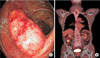
A 65-year-old woman was referred to Department of Surgery for treatment of an ascending colon cancer. A colonoscopy revealed a 2-cm-sized ulcerofungating mass in the cecum (Fig. 1A). A tumor biopsy indicated a moderately differentiated adenocarcinoma. An abdominal computed tomography scan showed an ulcerofungating mass in the cecum, and a radionuclide positron emission tomography scan of the torso (basal skull to proximal thigh) using 18F-fluorodeoxyglucose revealed a hypermetabolic lesion in the cecum (Fig. 1B). The patient's carcinoembryonic antigen levels were 3.52 ng/mL. We planned to perform a single-port plus an additional port robotic right colectomy using the Da Vinci Single-Site platform.
Technique
In this study, a totallty robotic, reduced-port surgical approach using the Da Vinci Single-Site platform and Endowrist system was employed. The surgery included lymphovascular dissection around the central vascular trunk, lateral and hepatic flexure mobilization, and intracorporeal side-to-side anastomosis using the robotic stapler
Installation and docking of robotic system
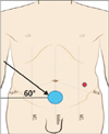
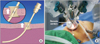
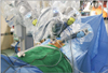
For bowel preparation, colonic lavage was performed the day before the surgery using 2 L Coolprep (TaeJoon Pharmaceuticals, Seoul, Korea). Systemic prophylactic antibiotics were administered intravenously at the time of general anesthesia induction. The surgery was performed under general anesthesia with the patient in the lithotomy position. A single 3.5-cm pfannenstiel incision was made and the peritoneum was opened. The Glove port (Nelis, Bucheon, Korea) was inserted into the intraabdominal space. After achieving pneumoperitoneum with insufflations of CO2 to 12 mmHg, a Da Vinci 8.5-mm endoscope with a 30° angled view was inserted. An additional 12-mm Stapler 45 Cannula with Stapler 45 Reducer (Intuitive Surgical, Sunnyvale, CA, USA) for the wristed robotic instruments was inserted in the left lower quadrant under direct visualization. Laparoscopic exploration was undertaken using laparoscopic instruments via the additional robotic port, and accessory lumen of the single-port. The patient was put in the Trendelenburg position at 30° and tilted left side down at an angle of 15°. For the initial exposure, the greater omentum was flipped over the transverse colon toward the liver, and small bowel loops were retracted into the left side of patient's abdominal cavity. The robotic cart was subsequently placed oblique to the surgical table at an angle of 60° (Fig. 2). The camera arm was docked to the corresponding trocar, and the robotic 3-dimensional laparoscope was reinserted and attached to the camera arm. The 5 × 250-mm curved cannulas were lubricated and inserted under direct visualization via the single-port, and the R1 and R2 arms were docked using the Dock Assistant Tool (Intuitive Surgical, Sunnyvale, CA, USA) to create space around the instrument arm during docking. The R3 arm was docked to the 12-mm Stapler 45 Cannula with Stapler 45 Reducer. To maximize the instrument arm's range of motion, the set up joints of the R1 and R2 arms were positioned in straight lines. Additionally, to secure the distance between the tip of instruments and surgical field, such as superior mesenteric vessels and retrocecal recess, remote centers of R1, R2 cannulas and camera port were lifted upward out of the abdominal wall (Fig. 3A), and all three remote centers were aligned at the same level (Fig. 3B). The R1 arm was used for the nonwristed semirigid Maryland Dissector available for bipolar coagulation, the R2 arm was used for the Cadiere Forcep, and the R3 arm was used for the wristed monopolar curved scissor (Fig. 4).
CME with central vascular ligation by medial to lateral approach
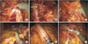
The robotic second arm of Da Vinci Single-Site platform was used to lift up the ileocolic pedicle, and dissection was commenced along a vertical line to expose the superior mesenteric vein with stable and durable retraction. Primary vascular control with a D3 lymphadenectomy at the origin of the ileocolic, right colic, and middle colic vessels was performed along the right border of the superior mesenteric vein. The ileocolic vessels were ligated using EndoWrist Clip Appliers (Intuitive Surgical) (Fig. 5A) and transected at the root, while the dissection continued upwards to the gastrocolic trunk. We transected the colonic branch of the gastrocolic trunk, preserving its pancreatic and gastric branches, and exposed the middle colic artery at its origin (Fig. 5B). Following lymph node dissection in this region, the middle colic artery was ligated (Fig. 5C). An avascular surgical plane composed of Toldt's line, and prerenal fascia was exposed to uncover the head of the pancreas, second portion of the duodenum, right gonadal vessels, and ureter. The integrity of the mesocolon was strictly preserved similar to total mesorectal excision for rectal cancer. The transverse mesocolon was suspended by the robotic second arm, and the lesser sac was entered just above the head of the pancreas. Lateral mobilization was performed by retracting the cecum anteriorly, medially, and superiorly with the Cadiere Forcep in the robotic second arm. The peritoneum at the retrocecal recess was incised, and retroperitoneal avascular plane was developed.
Hepatic flexure mobilization
The greater omentum of the transverse colon was subsequently transected using the EndoWrist One Vessel Sealer (Intuitive Surgical) and replaced with the monopolar curved scissors to allow entry into the lesser sac, joining the previous surgical plane of medial dissection (Fig. 5D). At this point, the fixed second arm traction on the greater omentum, and posterior wall of the stomach significantly facilitated the dissection of the omentum. After the lateral peritoneum of the ascending colon and the attachment of the hepatic flexure were detached, the gastrocolic ligament and right side of the greater omentum were dissected.
Intracorporeal anastomosis and specimen extraction
The transverse mesocolon and small bowel mesentery were divided to achieve anastomosis using the EndoWrist One Vessel Sealer. The transverse colon and terminal ileum were then transected using EndoWrist Stapler (Intuitive Surgical) through Stapler 45 Cannula. An enterotomy and colostomy were made by the monopolar curved scissors and some bowel contents were sucked by EndoWrist One Suction/Irrigator. EndoWrist Stapler was introduced to create a side-to-side anastomosis in an isoperistaltic manner (Fig. 5E). The monopolar curved scissors were replaced with Suture Cut Needle Driver (Intuitive Surgical), and the stapler insertion site was then closed with continuous stitches using V-Loc suture (Covidien, Mansfield, MA) (Fig. 5F). Finally, a drainage tube was placed at right paracolic sulci via the R3 trocar, and the specimen was wrapped in a sterile bag and extracted via the single-port incision.
Clinicopathologic outcomes
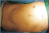
A single-port plus an additional port robotic approach facilitated lymphovascular dissection around the central vascular trunk and intracorporeal anastomosis with minimal external instrument conflicts. The procedure was successfully completed, and the total operative and docking times were 280 and 25 minutes, respectively. The surgery was uneventful, and conversion to conventional robotic or laparoscopic surgery was not required. Total incision length was 47 mm (Fig. 6). Passing of flatus took place on postoperative day 4, and the patient was discharged from hospital on postoperative day 8. The patient's pathology results indicated a moderately differentiated pT3N0 1.5 × 1.0-cm adenocarcinoma, with a 31-cm proximal margin and a 50-cm distal margin. No positive lymph node metastasis was found out of the total 36 pericolonic lymph nodes resected.
DISCUSSION
CME is the resection of the tumor by sharp dissection of the visceral plane from the parietal fascia layer, along with the entire regional mesocolon in an intact package. Hohenberger et al. [3] introduced a procedure with similar concept to the concept of "CME and central vascular ligation" for colonic cancer. They performed the procedure in an open approach, which reduced local recurrence and improved long-term cancer-related survival in accordance with the concept of total mesorectal excision for rectal cancer.
With minimally invasive surgery becoming increasingly common, efforts to further improve the cosmetic outcomes of surgery and reduce port-related morbidities of laparoscopic surgery, which include the size and number of ports, continue. The potential advantages of this approach are associated with improved cosmetic outcomes, less postoperative incisional pain, reduced risks of hemorrhage from port sites, fewer incisional hernias and organ injuries, and fewer wound complications. However, single-port laparoscopic CME for right-sided colon cancer is a challenging procedure, even in the hands of an experienced laparoscopic colorectal surgeon, because of the difficulties of creating triangulation, and the complex and variable vascular anatomy of the right colon.
Recently, the Da Vinci Single-Site platform, which was installed on the Da Vinci Si system, was introduced, and this system may help to overcome some of the difficulties of SPLS. The principal difference between the standard robotic approach and the new Single-Site platform is the improved triangulation, which makes surgical procedures using this platform easier than standard SPLS. On this platform, the 2 crisscrossed curved instruments are "repositioned" by the software to fit the corresponding hand of the surgeon on the console. However, this approach has not been widely used for colon cancer because of some limitations, including the absence of wrist function, which is usually recognized as one of the major advantages of the Da Vinci robotic system; other limitations include the limited range of motion of the semi rigid robotic instruments and limited availability of instruments for the Single-Site system. Morelli et al. [6] was the first to perform a right colectomy for a benign tumor using the Single-Site platform, and several authors [78] have reportedly performed a robotic colectomy using the Da Vinci Single-Site platform. However, there are some technical differences between the present study and those studies. First, in those reports, procedures were carried out using only Single-Site platform, whereas CME with meticulous dissection was performed using Single-Site platform and EndoWrist in our study. Second, Morelli et al. [6] performed hand-sewn ileo-colic anastomosis extracorporeally and Spinoglio et al. [7] performed stapled side-to-side anastomosis using laparoscopic linear staplers intracorporeally, we made side-to-side anastomosis using EndoWrist Stapler and wristed suturing intracorporeally.
Single-Site platform with various wristed robotic instruments via additional robotic port have some technical advantages including increased maneuverability of instruments and increased accuracy of anatomical dissection. These characteristics are especially important when precise lymph node dissection along the superior mesenteric trunk is required for D3 lymphadenectomy while minimizing blood loss. Additionally, this approach can provide safe intracorporeal anastomosis.
Although intracorporeal anastomosis has some advantages, including less complications related to the alignment of the mesentery and less dissection of distal transverse colon for a tension-free anastomosis, laparoscopic intracorporeal anastomosis is not widely applied. This is because intracorporeal suturing with laparoscopic instruments is challenging and takes a longer operation time than does extracorporeal anastomosis with an inexperienced surgeon. Bergamaschi et al. [9] reported short-term outcomes of 111 right colectomies with totally stapled intracorporeal anastomosis and commented that it can increase risk of inadvertent strictures caused by posterior bowel wall involvement during the stapling procedure. In addition, Hellan et al. [10] compared 57 extracorporeal anastomosis and 23 intracorporeal anastomosis in laparoscopic right hemicolectomy, and reported longer operative time in the intracorporeal anastomosis group. However, robotic intracorporeal anastomosis can be easily performed with safe microsuture and steady traction.
In this case, we lifted the remote centers of R1, R2 cannulas and camera port upward out of the abdominal wall to secure the surgical space between the tip of the instruments and the operative field because 250-mm curved cannulas are too long for the dissection around central vascular trunk. In our experience, although Single-Site port was designed for the Single-Site platform, lack of durability and occurrence of air-leaks was observed when the remote centers of the robotic ports were placed at the level out of the abdominal wall [8]. Once we changed the access port, these problems resolved.
In conclusion, single-port plus an additional port robotic surgery for right-sided CME and intracorporeal anastomosis appears to be feasible and safe. This system can overcome certain limitations of the previous robotic systems and conventional SPLS.
 XML Download