

 PDF
PDF ePub
ePub Citation
 Print
Print


INTRODUCTION
Multiple endocrine neoplasia type 2A (MEN 2A) is an autosomal dominant disease that is characterized by medullary thyroid cancer, pheochromocytoma, and primary hyperparathyroidism. It is a rare disease, with a prevalence of 2.5 per 100,000 people, and it is caused by a germ-line mutation in the rearranged during transfection (RET) proto-oncogene on chromosome 10 [1]. It mainly presents as medullary thyroid cancer followed by pheochromocytoma. Primary hyperparathyroidism occurs in about 20%–30% of patients with MEN 2A, which is less than the prevalence of medullary thyroid cancer or pheochromocytoma; in addition, its symptoms and signs are usually mild [123].
The autotransplantation of parathyroid tissue is a useful technique for preventing hypoparathyroidism during thyroidectomy or parathyroidectomy, and it was first described in animal experimentation by Halsted in 1909 [4]. When parathyroid glands are injured during thyroid surgery or when a patient requires total or subtotal parathyroidectomy for hyperparathyroidism, some parathyroid tissue is autotransplanted in the sternocleidomastoid muscle or the brachioradialis muscle. The success rate is about 75%–100% [5]. Rarely, patients experience hypoparathyroidism due to failure of autotransplantation or hyperparathyroidism due to the proliferation of autotransplanted tissues.
CASE REPORT
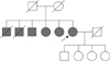
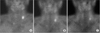
A 68-year-old female with MEN 2A had a family history of thyroidectomy for medullary thyroid cancer (Fig. 1). In addition, 15 years prior, she underwent left adrenalectomy for pheochromocytoma; 6 years prior, she underwent total thyroidectomy, central and right lateral neck lymph node dissection, and subtotal parathyroidectomy with autotransplantation in the left sternocleidomastoid muscle for medullary thyroid cancer and primary hyperparathyroidism. At this time, the serum calcium level was 10.7 mg/dL (normal range, 8.6–10.2 mg/dL) and the parathyroid glands were enlarged bilaterally; the intact parathyroid hormone (iPTH) level was not determined. After the surgery, follow-up serum calcium and iPTH levels were within the normal ranges. Five years later, her iPTH level increased (113.10 pg/mL; normal range, 15–65 pg/mL), and she experienced hypercalcemia (11.1 mg/dL) with elevated iPTH (122.30 pg/mL) in the next year. On ultrasonography, a 1.3 × 0.6-cm-sized ovoid suspicious mass was detected in the left sternocleidomastoid muscle on the level 3 or 4 (Fig. 2). A core needle biopsy on the mass confirmed it to be parathyroid tissue. On additional sestamibi scan, a focal hot uptake was observed at the left neck (Fig. 3). Therefore, she underwent mass excision. A 1.6-cm-sized, well-circumscribed mass was detected in the sternal part of the left sternocleidomastoid muscle, excised (Fig. 4), and histologically confirmed to be parathyroid adenoma.
DISCUSSION
The main objective of surgery for hyperparathyroidism is the correction of the hyperparathyroidism while minimizing hypoparathyroidism and recurrent postoperative hyperparathyroidism. Therefore, the extent of parathyroidectomy must be considered carefully. MEN 1 usually presents primary hyperparathyroidism (>90%), and it is also common for it to persist or recur after parathyroidectomy. Thus, the initial surgical approach of choice in MEN 1 patients with hyperparathyroidism is either subtotal or total parathyroidectomy with autotransplantation of excised pararthyroid tissue. The rate of hyperparathyroidism in MEN 2A is less common (20%–30%) and the symptoms and signs of hyperparathyroidism are usually milder than it is in MEN 1; therefore, the parathyroidectomy in MEN 2A patients with hyperparathyroidism can be less aggressive than in MEN 1 patients with hyperparathyroidism, although there is some debate [23].
Several studies on preserving parathyroid glands in MEN 2A have been performed. Yoshida et al. [6] reported long-term postoperative follow-up of serum iPTH level in 12 MEN 2A patients who underwent total parathyroidectomy with autotransplantation, along with surgery for medullary thyroid cancer. These patients maintained normal iPTH level without hypoparathyroidism, suggesting that total parathyroidectomy with autotransplantation is a feasible management method for preventing hyperparathyroidism in MEN 2A. In this case, however, although grossly normal parathyroid tissue was autotransplanted, it recurred as a parathyroid adenoma 6 years later. Thus, it appears that parathyroid tissue in MEN 2A retains the proliferative capacity to form adenoma or hyperplasia, even in cases of autotransplantation.
The recurrence rate after autotransplantation of parathyroid tissue is 7%–30%, and the risk of recurrence is higher when associated with genetic diseases such as MEN [7]. Transition or transversion of the 634 codon in the RET proto-oncogene is the main gene mutation seen in MEN 2A [3]. Mulligan et al. [8] reported that 73% of MEN 2A patients with hyperparathyroidism had a mutation that changed the cysteine residue in the 634 codon to arginine (C634R, TGC>CGC). This mutation may modify parathyroid tissue in patients with MEN 2A to be more proliferative than normal, increasing the risk of recurrence. Hedback and Oden [9] reported that the postoperative recurrence rate for hyperparathyroidism patients was 7% during a mean of 10 years of follow-up, and a recurrence occurred 33 years after surgery, suggesting that long-term follow-up is necessary in such patients.
We usually autotransplant parathyroid tissue in the sternocleidomastoid muscle, but this may require general anesthesia for re-excision when it recurs as a parathyroid adenoma or hyperplasia; therefore, the adverse effects of general anesthesia need to be considered. Transplantation in the brachioradialis muscle is safer, as local anesthesia may be appropriate for reexcision of recurrent parathyroid adenoma or hyperplasia. Furthermore, Cavallaro et al. [10] reported that parathyroid autotransplantation in the forearm subcutaneous tissue had a good recovery rate of graft function (96% at 3 months after surgery), and it is easy and safe to control graft function. Therefore, autotransplantation in the brachioradialis muscle or in the forearm subcutaneous tissue may be preferred for patients who are at high risk for general anesthesia complications due to old age or cardiopulmonary disease.
Compared to patients with MEN 1, hyperparathyroidism in MEN 2A patients is less frequent and carries less severe symptoms. However, we identified the potential of recurrent hyperparathyroidism in MEN 2A patients in this case. Thus, long-term regular monitoring of serum calcium and iPTH levels is necessary after parathyroid autotransplantation.


 XML Download