

 PDF
PDF ePub
ePub Citation
 Print
Print


INTRODUCTION
An estimated 185 million individuals are chronically infected with HCV worldwide [1]. Of all HCV-infected individuals, 20%-30% develop liver cirrhosis and 1%-4% of all patients with liver cirrhosis develop hepatocellular carcinomas (HCC) [2]. HCV infection is the most common indication for liver transplantation (LT) in Western countries. In Korea, 1%-2% of the population is infected with HCV, and 15%-20% of these infected individuals have chronic liver diseases related to HCV infection [34]. As the prevalence has increased, HCV-related cirrhosis and HCV-related HCC will gradually become more common indications for LT in Korea [5].
Although LT offers the optimal treatment for HCV-related end-stage liver disease and HCC, graft reinfection with HCV is not acute, but rather immediate and universal in all patients who are HCV RNA-positive at transplantation [6]. HCV RNA levels increase when immunosuppression is the highest during the first few months after transplantation. The progression of fibrosis in LT patients is accelerated compared to that in nontransplanted patients because the virus is more aggressive after LT than it is in immunocompetent subjects.
Genetic variation in interleukin-28B (IL28B) predicts hepatitis C treatment-induced viral clearance. Single nucleotide polymorphisms in IL28B have varied distributions among ethnic groups. East Asian populations such as those in Korea, Japan, and China have the highest frequencies of single nucleotide polymorphisms in alleles associated with HCV clearance [7].
The cumulative number of HCV-related cirrhosis and HCV-related HCC cases in Korea is very small; therefore, we collected data of LT recipients with HCV from three major centers. We conducted a survey of HCV RNA-positive patients who underwent LT and investigated the prognostic factors for patient survival.
METHODS
Patients
This was a multicenter study involving three LT centers in Korea: Samsung Medical Center (SMC), Asan Medical Center (AMC), and Seoul National University Hospital (SNUH). We did not consent from patients for usage of their clinical records in this study because of retrospective study. Each center's Institutional Review Board (IRB) approved this study (SMC IRB No. 2014-07-031, AMC IRB No. S2015-1341-0003, SNUH IRB No. 1407-139-597) because written informed consents were not given by patients. We retrospectively evaluated patients undergoing their first LT between 1994 and 2012. Data from all consecutive HCV RNA- positive cases were reviewed during this period. Each institution utilized a survey with study questionnaire items. The information and/or records of patients de-identified prior to analysis.
HCV RNA quantitation was assessed using the quantitative-branched DNA amplification assay. HCV genotyping was performed following the standard method using reverse hybridization assays after amplification with a polymerase chain reaction assay, based on Simmonds' classification.
Among the 255 cases with HCV-related cirrhosis who underwent LT during the study period, 63 cases were excluded due to re-transplantation (n = 13), missing results of HCVRNA (n = 23), and HCV-RNA negativity (n = 27). Among the remaining 192 included patients, we identified the causes for graft failure and mortality. We investigated the risk factors associated with patient survival, but exclude hospital mortality (n = 23) for the identification of risk factors which were related with mortality in stable liver transplant recipients with more than three months after transplantation.
Evaluated variables
The following variables were obtained from the medical record review in response to the survey. Collected recipient factors included patient age, gender, pre- and posttransplant antiviral treatment, HCV genotype, model for end-stage liver disease (MELD) score, the coexistence of HCC, the coexistence of HBV or human immunodeficiency virus (HIV), antiviral treatments received after transplantation, the type of calcineurin inhibitor received, the use of mycophenolate mofetil, steroid withdrawal, biopsy-proven acute rejection, HCV recurrence, the response to antiviral treatments, and the outcomes. Additionally, donor age, ischemic time, and the type of partial liver graft were added as variables. Finally, we recorded information on patient survival and calculated time to death. However, we did not incorporate into the analysis any other incomplete variables that may be associated with patient survival, such as IL-28 gene polymorphisms, histological findings, biliary complications, and infectious episodes.
The diagnosis of acute rejection followed internationally accepted histologic criteria (Banff guidelines) based on liver biopsies [8]. HCV recurrence was diagnosed based on histology, biochemistry, and/or the detection of HCV RNA in the serum.
Statistical analysis
Continuous data are expressed as the median and range and were compared using the Mann-Whitney U test. Categorical variables are reported as numbers (proportions). Comparisons between groups for categorical data were performed using the chi-square test or Fisher exact test, when appropriate. Patient survival rates were evaluated using the Kaplan-Meier method and compared using the log-rank test. Clinical variables found to have prognostic significance by univariate analysis were entered into a Cox multivariate proportional hazards model to determine factors that independently predict patient survival. The cutoff values for the continuous variables were set according to each receiver operating characteristic (ROC) curve. Statistical significance was set at a P-value of less than 0.05. Statistical analysis was performed with IBM SPSS Statistics ver. 21.0 (IBM Co., Armonk, NY, USA).
RESULTS
Patient survival and outcomes
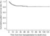
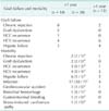
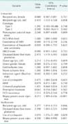
The 1-, 3-, and 5-year cumulative patient survival rates were 78.8%, 75.3%, and 73.1%, respectively (Fig. 1). The causes of graft failure and mortality are summarized in Table 1. Thirty patients (15.6%) developed graft failure during the observation period and 13 patients underwent retransplantation. Fifty patients (26.0%) died during the observation period. Most cases of mortality (38 of 50, 76%) occurred less than one year after transplantation. The patient survival curve showed an abrupt decrease, but then stable survival after two years after transplantation and onward. Recurrent HCV infection and hepatic failure (n = 17) and chronic rejection (n = 10) were the causes of graft failure. Chronic rejection was the main cause of graft failure in patients less than one year after transplantation. Infection (n = 21) and recurrent HCV infection with hepatic failure (n = 15) were the leading causes of recipient death. Infection was the main cause of hospital mortality in patients one year after transplantation.
Baseline characteristics
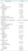
Among the 192 patients that were identified, we investigated the 169 patients, who represented all patients except those who died in the hospital (n = 23). The characteristics of the 169 HCV RNA-positive LT recipients compared in this study are summarized in Table 2. There were 118 men and 51 women, with a median age of 56 years (range, 34-71 years). The median follow-up period was 38 months (range, 1-157 months), with a wide spectrum of follow-up duration due to death or shorter observation period from LT. The median MELD score and median HCV RNA levels were 16 (range, 6-50) and 133,568 IU/mL (range, 12-26,000,000 IU/mL), respectively. One hundred eleven patients (65.7%) had HCV genotype 1 and 42 patients (24.9%) had HCV genotype 2. The number of patients with coexisting HBV infection, HIV infection, and HCC was 21 (12.4%), 1 (0.6%), and 77 (45.6%), respectively. There were 135 living donor liver transplantations (LDLTs) (79.9%) and 34 deceased donor liver transplantations (DDLTs) (20.1%). The median age of the donors was 32 years (range, 16-70 years), and the graft type in the living donors was the right liver in 125 patients (74.0%). The median cold ischemic time and median warm ischemic time were 81 and 39 minutes, respectively.
Prognostic factors for patient survival
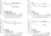
Recipient and donor factors were analyzed for their association with overall mortality. The results of the univariate and multivariate analyses are shown in Table 3. The univariate analysis revealed that recipient age ≥ 60 years (P = 0.018), HCV RNA levels at pretransplant (P = 0.023), DDLT (P = 0.020), donor age ≥ 30 years (P = 0.019), the use of cyclosporine (P = 0.025), and biopsy-proven acute rejection (P < 0.001) were significant predictors of poor outcome in HCV RNA-positive recipients. The duration of steroid use did not affect patient survival. The ROC curve did not reveal a significant cut-off value for HCV-RNA levels in terms of patient survival. The multivariate analysis showed that recipient age ≥ 60 years (P = 0.046), DDLT (P = 0.040), the use of cyclosporine (P = 0.029), and biopsy-proven acute rejection (P = 0.001) were independent prognostic factors for mortality. The Kaplan-Meier survival curves stratified by these factors are presented in Fig. 2.
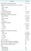
Antiviral treatments in pre- and posttransplant
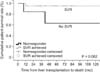
A summary of the antiviral treatments is shown in Table 4. Of the 169 recipients, 129 did not receive antiviral treatment in the pretransplant period and 30 underwent antiviral treatment. After LT, 75 patients received universal prophylaxis and 15 patients underwent preemptive treatment due to HCV reactivation. Most patients did not undergo a protocol biopsy, and HCV-RNA levels were monitored at every visit. HCV recurrence was detected in 97 patients (57.4%). Among the 97 patients with HCV recurrence, 48 patients were treated with antiviral therapy. The survival rates were higher in patients with sustained viral response (SVR) than in patients without SVR, but there was no statistically significant difference in patient survival between the two groups (P = 0.062) (Fig. 3).
DISCUSSION
Literature from the United Network for Organ Sharing (UNOS) database reported a 5-year patient survival rate of 76%, and a study from the European Liver Transplant Registry (ELTR) reported a 5-year patient survival rate of 65% [910]. Recently, nationwide survey in Japan of LDLT reported a 5-year patient survival rate of 72% [11]. Our study here is the largest case series of LT for HCV RNA-positive recipients in Korea. A total of 192 recipients from three large institutions were reviewed and found to have a 5-year patient survival rate of 73.1%. Based on these studies, the outcomes of the present study are similar to that of the ELTR, UNOS, and Japanese survey. Comparisons of the survival rates of HCV recipients between studies should be interpreted with caution because our study excluded patients with operative mortality, hospital mortality, and retransplantation. The proportion of IL28B in Korea is higher than in European countries [7], however survival rates of Korea were similar to other countries despite advanced surgical techniques and perioperative managements in LT.
We selected liver transplant recipients with HCV-RNA positive in the pretransplant period because there have been no reports of posttransplant HCV recurrence in HCV RNA-negative recipients. Antiviral therapies based on Pegylated interferon-alpha (PEG-IFNα) and ribavirin (RBV) have been used to treat HCV in decompensated patients on the transplant waiting list until they are HCV RNA-negative [12]. However, this therapy is limited due to poor tolerance, poor efficacy, and serious adverse events seen in those waiting for LT [13].
Posttransplant viral load is an important marker of disease severity, while pretransplant viral load predicts more severe HCV recurrence after transplantation [14]. Negative HCV viral load at the time of transplantation does not preclude HCV recurrence in the liver graft. A peak posttransplant HCV viral load >107 IU/mL was an independent predictor of graft loss and mortality [15]. Present study showed that prognostic factors, including recipient age > 60 years, DDLT, the use of cyclosporin, and biopsy-proven acute rejection were closely associated with patient mortality. High viral load was associated with mortality in univariate analysis, but a cutoff value for the HCV RNA level was not drawn. Universal prophylaxis should be initiated soon after LT because the viral load is at its lowest level and fibrosis in the graft is absent [16]. However, antiviral therapy may be less effective in the early posttransplant period secondary to strong immunosuppression, and tolerance is low because of the high risk of poor hematological tolerance, acute rejection, and sepsis [1718].
Posttransplant patients with HCV recurrence have significantly diminished survival compared to posttransplant patients with no recurrence. The progression of recurrent HCV is variable and the key risk factors remain unclear. Many factors have been reported to play a role prior to LT (genotype 1, viral load, and female gender) or after LT (time of cold or warm ischemia, blood transfusions, steatosis in the liver graft, age of the donor, the use of antilymphocytes, and coinfection with HIV) [1319]. The early detection of HCV recurrence is crucial because HCV-infected patients appear to respond better to early antiviral therapy [19]. The current clinical standard for early detection is for protocol liver biopsies to be performed every 1-2 years after LT, as HCV-infected recipients are at increased risk of HCV-mediated graft cirrhosis [20]. Antiviral treatment is delayed until there is histological evidence of recurrent hepatitis in many transplantation centers. However, none of the centers in the present study performed these protocol biopsies.
The successful treatment of recurrent HCV, which is demonstrated by sustained HCV clearance or an SVR, is associated with reduced liver-related mortality and improved overall survival. The combination of PEG-IFNα and RBV is the current standard of care [1321]. Our study also revealed this effect in the SVR group in recurrent HCV patients, but this did not reach statistical significance. However, the PHOENIX trial of PEG-IFNα and RBV given preemptively after the transplant for HCV found no clear benefits when considered in the context of side effects [18]. In a very small study, donor or recipient IL28B genotypes were shown to predict SVR with PEG-IFNα and RBV therapy, and IL28B status was related to SVR after LT [22]. However, this effect of the IL28B genotype was not identified in the present study, and our study did not reveal an association between IL28B and patient survival.
The findings of this retrospective, multicenter study are limited by several factors inherent to the study type, including variability in documentation, differences in the selection criteria and data collection, and missing data. To minimize variability, we sent a standardized collection form containing 56 questions to the transplant centers. The answers were either multiple-choice or involved providing a name or a specific value. However, the quality of the pretransplant interviews from which the baseline data were derived, and the quality of the posttransplant follow-up data across the three centers may have varied. Furthermore, subjects had varying follow-up durations. We did not have data on the onset of biopsy-proven acute rejection or the date of graft failure. To address these limitations, a well-designed prospective study is needed.
In conclusion, this retrospective analysis of the largest three LT centers for HCV RNA-positive recipients in Korea revealed 1-, 3-, and 5-year survival rates of 78.8%, 75.3%, and 73.1%, respectively. The prognostic factors for patient survival except hospital mortality revealed that recipient age > 60 years old, DDLT, the use of cyclosporin, and biopsy-proven acute rejection are closely associated with patient mortality. Present study revealed that patient survival rates in HCV patients after LT in Korea were comparable other countries.
 XML Download