

 PDF
PDF ePub
ePub Citation
 Print
Print


INTRODUCTION
Choledochal cysts are rare hepatobiliary abnormalities with an incidence of 1 per 100,000–150,000 in the general population and 1 in 1,000 in Asia. The widely accepted Todani classification describes 5 types of choledochal cyst [1]. However, isolated cysts of the cystic duct are extremely rare and not included in the Todani classification; some authors refer to them as "type VI," whereas others consider them a subtype of Todani type II [234]. Here, we present our experience with an unusual isolated cystic duct cyst with associated with stones in a 4-month-old infant.
CASE REPORT
A 4-month-old male patient presented to the pediatric emergency room after 2 days of abdominal discomfort and vomiting. He had been born healthy at a gestational age of 40 weeks and a weight of 3.7 kg. At 3 months old, he had visited his local clinic for evaluation and management of enteritis. Abdominal ultrasonography (USG) had incidentally revealed wall thickening and edema of the gallbladder (GB). However, he had shown no symptoms of GB abnormality, and his blood test results had been normal, so the lesion had not been treated immediately. One month later, he vomited greenish material at home and showed signs of abdominal pain, whereupon he was admitted to our hospital.
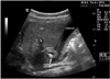
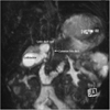
Follow-up abdominal USG revealed acute GB inflammation with stones and sludge (Fig. 1). Magnetic resonance cholangiopancreatography (MRCP) showed fusiform dilatation of the cystic duct with internal sludge and stones, diffuse GB wall thickening, and mild distention without evidence of anomalous pancreaticobiliary ductal union (Fig. 2). Liver enzymes were normal with leukocyte count 7,580/µL and C-reactive protein 0.07 mg/dL, respectively.
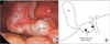
We initially decided to treat the patient with antibiotics (intravenous cefotaxime 425 mg, every 8 hours during breast-feeding), expecting improvement in the symptoms and USG findings. However, the follow-up USG after 1 week showed no significant improvement, so we decided to perform a laparoscopic cholecystectomy. Laparoscopy revealed an isolated cystic duct cyst (Fig. 3A), and GB aspiration revealed that the bile was clear. It was very difficult to dissect between the cyst and the common bile duct (CBD) owing to the severe inflammation caused by the impacted stones; we therefore decided to switch to a simple open cholecystectomy for safety. The GB and cyst were successfully excised without need for further complex procedures. Two stones were found, one in the cyst and one in the distal outlet of the cystic duct (Fig. 3B). Cystic duct dilatation was confirmed grossly with internal diameter of 1.8 cm in average in the pathology report. The postoperative course was uneventful, and the patient was discharged on the seventh postoperative day.
DISCUSSION
Isolated cystic duct cysts are very rare; with only around 20 cases previously reported in the literature, their incidence cannot be reasonably estimated [2345678]. Serena Serradel et al. [4] and Bode and Aust [2] initially misdiagnosed such cysts as Todani type II choledochal cysts, and Loke et al. [3] also mentioned that the case they encountered resembled a type II cyst. Isolated cystic duct cysts can be treated by simple cholecystectomy, similarly to type II cysts that do not arise from the intrapancreatic portion of the CBD [9]. However, we believe that isolated cystic duct cysts should be classified separately from other types of choledochal cysts, for instance as "type VI" (the term used by Serena Serradel et al. [4]); this will help clarify the nature of such lesions and help establish proper management methods.
The most common symptoms of cystic duct cysts are pain in the upper abdomen [45678] or jaundice [3]; our patient's main complaints were vomiting and abdominal pain. The wall of the cyst was very thin and weak and could easily have been perforated. Nevertheless, GB aspiration showed our patient's bile to be clear rather than the normal green color, suggesting obstructed communication between the GB and the CBD. This disconnection may have developed as a result of obstruction of the cystic duct lumen owing to disruption of the mucosal layer by recurrent inflammation and impacted stones. The obstruction, in turn, may have caused the observed wall thickening and edema of the GB, resulting in secondary inflammation. Overall, the clinical presentation of this case resembled that of Mirizzi's syndrome.
As mentioned in earlier studies, radiological imaging is important to ensure correct diagnosis and an appropriate treatment plan. Abdominal USG is a good initial screening method [10]. Endoscopic retrograde cholangiopancreatography (ERCP) can reveal the biliary anatomy in detail, including any sign of an abnormal pancreatobiliary junction, and MRCP, a less invasive method of evaluating the biliary tree, is also highly effective. We used serial USG and MRCP to diagnose our patient's condition and check whether the antibiotics had had an effect.
As recommended in previous reports of isolated cystic duct cysts [24], our patient underwent cholecystectomy with excision of the cyst and transection of the cystic duct preserving normal anatomy of CBD. However, the necessary extent of the surgery may vary depending on the length of distal cystic duct between the cyst and CBD or on the diameter of the cystic duct junction with the CBD; in the cases reported by Loke et al. [3] and Shah et al. [8] the junction had a wide diameter, so those authors performed Roux-en-Y hepaticojejunostomies. Bresciani et al. [5] and Conway et al. [6] were able to apply minimally invasive approaches because, in their cases, the cyst was easily dissected, and the diameter of the distal cystic duct was narrow enough to ligate laparoscopically.
We have reported an unusual case of an isolated cystic duct cyst associated with gallstones. Proper diagnosis based on radiological imaging and complete surgical excision of the GB and cyst are keys to the treatment of such cysts. Once the diagnosis is made, surgical treatment should not be delayed. To achieve better understanding of this type of extremely rare biliary tract anomaly, cases like this should be reported more and included as a separate category in classifications of choledochal cysts.
 XML Download