

 PDF
PDF ePub
ePub Citation
 Print
Print


INTRODUCTION
Breast cancer is increasing each year in the Korean population. Breast cancer is diagnosed by physical examination, mammography or ultrasound and confirmed by biopsy [1]. Patients with palpable breast mass or breast mass identified by ultrasound is not difficult to diagnose. However, nonpalpable breast lesions or microcalcification, which are identified only in mammography, are difficult to diagnose. Wire localization biopsy using a hook wire has been widely used for those lesions [2]. In recent years, stereotactic vacuum-assisted breast biopsy (VAB) was introduced for accurate and even minimally invasive methods [34].
Stereotactic VAB is usually performed in the prone position, which requires expensive and specially dedicated tables. Stereotactic VAB in the upright position overcomes these disadvantages, and the procedure is easier and faster. However, maintaining this position is difficult and the procedure is performed facing the patient, which causes anxiety and vasovagal syncope. Therefore, lateral decubitus positioning stereotactic VAB was introduced to compensate for these problems. We evaluated the usefulness of lateral decubitus positioning stereotactic VAB in this study.
METHODS
Between January 2009 and December 2014, we performed and evaluated stereotactic VAB under lateral decubitus position on 106 lesions that were clustered microcalcifications in mammography requiring biopsy in our Breast Center. Lateral decubitus positioning stereotactic VAB was performed on all of these patients using the 8G probe (Mammotome; Ethicon Endo-Surgery, Cincinnati, OH, USA). Specimen mammography was obtained to confirm microcalcification.
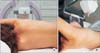
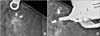
The patient lay on a stereotactic VAB table in the lateral decubitus position with the affected breast side up (Fig. 1A). The breast was compressed and fixed to the appropriate direction (Fig. 1B). First, we checked the mammography. An additional 15° paired stereotactic mammography was obtained. Then the computer program calculated the three-dimensional location, the horizontal (x) and vertical (y) distance and the depth (z) from the zero point of the stereotactic VAB device (Fig. 2).
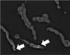
The attached needle was moved to the skin incision site we measured. Local anesthesia was done through the breast parenchyma under aseptic conditions. After the skin incision within 4 mm, the 8G VAB probe was inserted through the incision site. Stereotactic 15° paired mammographies were checked to find the presence of dislocation due to lidocaine infection or needle insertion. We obtained a specimen after the needle location was confirmed by checking the image (Fig. 3). One or two tissue specimens were collected from each of the six clockwise positions (2, 4, 6, 8, 10, and 12). If the calcification is still identified on stereotactic mammography or was not identified in the specimen mammography, additional sampling was done. The needle was removed from the biopsy site when the procedure was successfully completed. The biopsy site was manually compressed. Skin closure with a piece of strip (Steri-Strip; 3M, St. Paul, MN, USA) and a compression bandage were used for hemostasis. These procedures were all performed by a single physician.
RESULTS
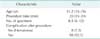
The average of patient age was 51.2 years (range, 35-76 years) and the average of procedure time was 20 minutes (range, 15-24 minutes). The average number of obtained specimens was 8.5 pieces (range, 6-12 pieces) (Table 1).
Mammography findings were distributed from category 3 to category 4 according to the breast imaging-reporting and data system (BI-RADS) classification. Microcalcification was observed in all specimen mammographies that underwent stereotactic VAB in 106 patients. The histological results for 10 patients (9.5%) were ductal carcinoma in situ (DCIS), 8 patients (7.5%) were focal atypical ductal hyperplasia (ADH), and 88 patients (83%) were benign (Table 2). There was neither vasovagal syncope event nor major complication. Minor hematoma was reported in 8 patients (7.5%).
Additional breast conserving surgery was performed for the 10 patients who were diagnosed with DCIS.
Additional wide excision was done for 2 of 8 patients diagnosed with ADH, due to diffusely scattered microcalcification. The final pathologic diagnosis was reported as ADH only. After discussion with the patients, we decided close follow-up for 2 patients and another 6 patients diagnosed with ADH. Also, 88 patients diagnosed with fibrocystic change were followed up 6 months later by mammography without additional surgery. They showed no interval change.
DISCUSSION
Traditionally, wire guided excisional biopsy is performed for non-palpable breast lesion and mammographically detected microcalcification. However, this method is more invasive, leaves a larger scar, is more complicated, and more time-consuming than the percutaneous biopsy method. Recently, stereotactic VAB was introduced for breast lesions like these. Many biopsy tools have been developed depending on the procedural approach and the position of the patient.
Stereotactic VAB can be classified as prone position [4567], upright position [89], and lateral decubitus position method.
In the prone position, patients feel more safe. But a special table for the prone position is expensive and more space is required. It is difficult to perform a procedure at this table for patients who have small breasts such as Asian women. Also, it is impossible for lesions close to the chest wall.
The upright position method compensates for these disadvantages. This method is more suitable for the average Asian woman who have small breast sizes. However, it is very difficult to fix the breast and maintain a stable position. Moreover, the procedure is performed in front of the patient; this causes anxiety to the patient, back pain and vasovagal reaction.
To compensate for the disadvantages of the prone or upright-positioning for stereotactic VAB, a technique using an add-on stereotactic unit with the patient in the decubitus position is used increasingly to avoid patient movement and syncope [10]. Recently, the decubitus table (DBI table, Medical Positioning Inc., Kansas City, MO, USA) has been developed and used with add-on stereotactic units.
Laterally decubitus position was introduced to overcome the problems of these two methods. In particular, it is possible to obtain more specimens by using an 8G probe instead of an 11G probe, for more accurate and faster biopsy [11]. In our study, an expensive prone table was not necessary during stereotactic VAB and its lack did not cause vasovagal reaction.
In our center, stereotactic VAB has been used for biopsy of microcalcification since 2009. However, the procedure cost is still more expensive than surgical excision. When the cost was burdensome for the patient, surgical excision using a hook wire was done. Also, patients with breast parenchyma lesser than 3 mm, making the procedure impossible, had surgical excision.
According to BI-RADS, 90 patients (84.9%) were classified as category 4, which was the majority among the calcified lesions confirmed by biopsy. There were biopsy cases with category 3 patients, which were microcalcifications increasing during their follow-up or patients who were anxious about the microcalcification itself. In our clinic, there was no case diagnosed as malignancy in category 3 lesions, making the category an important factor to diagnosis before biopsy is done. However, there were some cases diagnosed as malignancy. They were classified as category 3 in mammography, but category 4 in ultrasound showing a mass lesion. Therefore, BI-RADS classification is important in microcalcification lesions with no mass lesion shown in ultrasound.
In our study, there was no case diagnosed as malignancy in category 3, but there are some reports that the cancer rate of category 3 lesion is more than 20%. Therefore, biopsy is needed even in category 3 lesions in clinically needed cases. For this reason, one radiologist and one breast surgeon judge the necessity of biopsy in these cases in our clinic. Biopsy is done when any clinician declares the necessity of biopsy.
Stereotactic VAB can focus precisely on clustered small lesions. Therefore, this procedure is a very excellent way to obtain a minimum of specimens quickly. However, it is possible to obtain only the specimen around the needle in distributed lesions. Also, VAB cannot function well in a patient whose breast parenchyma is thin. In recent years, there are a lot of solutions to these problems, but it is still difficult to perform a biopsy in lesions close to the chest wall or skin.
The complication rate of VAB is reported as approximately 1%-4%, but serious complications requiring treatment are rare [1112]. In our study, there were only four cases of mild hematoma without any serious complication.
In conclusion, lateral decubitus position stereotactic VAB using 8G probe does not need a dedicated table, and is easier to maintain the position compared to upright stereotactic VAB. It can also aid in obtaining a large size of specimen, reducing the number of trials and procedure time. This makes lateral decubitus positioning stereotactic VAB an accurate, safe and simple biopsy method; and also reduces the patient's vasovagal reaction.
Our report has limitations such as the small number of cases with a short-term follow up of four years. Long-term research of the false negative rate in stereotactic VAB is needed in category 4 patients who have been diagnosed with a benign lesion, because they can increase in size or possibly form a mass. Thus, further improvements to the limitations our research can provide greater benefit to patients with microcalcification.

 XML Download