

 PDF
PDF ePub
ePub Citation
 Print
Print


INTRODUCTION
A duodenal stump fistula (DSF) forming after a gastrectomy for gastric cancer is a life-threatening complication. Even though the overall incidence is low (1.8%–3%), the DSF-related mortality rate is reported to be 7% to 67% [123]. There are many DSF-related complications leading to longer hospitalization times, such as intra-abdominal abscesses, wound infections, diffuse peritonitis, sepsis, malnutrition, pancreatitis, abdominal bleeding, and pneumonia [1].
DSF is thought to be caused by many factors such as inadequate closure of the duodenal stump, devascularization, cancer involvement or resection, an inflamed duodenal wall, local hematoma, incorrect drain position and postoperative distension of the duodenum [4]. Orsenigo et al. [5] first reported the risk factors associated with postoperative DSF to be heart disease, liver cirrhosis, intraoperative blood loss (>300 mL) and the absence of manual reinforcement. However, that analysis focused on intraoperative factors. The aim of the present study was to analyze the risk factors for DSF that could be revealed during the preoperative evaluation for obtaining informed consent before surgery. By identifying them, we should be able to pay more attention to patients who are in high-risk groups during the surgery and postoperative management. The risk factors for DSF after gastrectomy for gastric cancer were analyzed retrospectively, and we also describe the methods used for prevention and management of DSF in our institution.
METHODS
The records of 1,018 consecutive patients who underwent curative gastrectomy from November 2008 to December 2013 were reviewed retrospectively. All of the patients had gastric cancer. Among them, 716 had a duodenal stump after gastrectomy. The methods used for intestinal reconstruction were Billroth II gastrojejunostomy (B-II) with jejunojejunostomy (Braun's anastomosis) or Roux-en Y (R-Y) gastrojejunostomy for subtotal gastrectomy and R-Y esophagojejunostomy for total gastrectomy.
DSF was diagnosed by the presence of duodenal fluid in the surgical drainage and confirmed by a CT scan when needed. The location of any tumor in the stomach was classified into 4 regions: antrum, stomach angle to lower body, mid body to high body, and cardia/fundus [6].
Surgical procedures
Duodenal transection was performed using a linear stapler (DST Series TA 60 mm, Covidien, Boulder, CO, USA) in cases involving open gastrectomy. In laparoscopic cases, a laparoscopic linear stapler (ECHELON FLEX ENDOPATH, Ethicon Endo-Surgery, Cincinnati, OH, USA) was used for duodenal transection. For surgery involving B-II with Braun's anastomosis, a side-to-side gastrojejunostomy was made approximately 40 cm distal to the ligament of Treitz via the antecolic pathway, and Braun's anastomosis was performed about 25 cm distal to the gastrojejunostomy. In patients who underwent an R-Y anastomosis, the jejunum was brought through the antecolic route after its division. The distance between gastrojejunostomy and the jejunojejunal anastomosis was about 25 cm, which is considered adequate for preventing bile reflux, with respect to intestinal limb length and Roux stasis or kinking.
To prevent DSF during surgery, we used an absorbable reinforcement felt (Neoveil, Gunze Limited, Shenzhen, China) and a fibrin sealant (Tisseel, Baxter AG, Wien, Austria) on the duodenal stump.
Statistical analysis
All data were analyzed using IBM SPSS Statistics ver. 21.0 (IBM Co., Armonk, NY, USA). Categorical outcomes were analyzed using the chi-square test and Fisher exact test. Mean differences between the two groups (with and without DSF) were analyzed using Student t-test. Independent risk factors associated with DSF after gastrectomy were analyzed using logistic regression analysis. The odds ratios (ORs) were estimated with 95% confidence intervals (CIs). Differences were considered statistically significant at P < 0.05.
RESULTS
Demographic characteristics
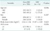
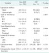
A DSF after gastrectomy for gastric cancer was found in 16 of the 716 patients (2.2%). The demographic characteristics of patients with and without DSF are summarized in Table 1. The 2 groups were similar with regard to age, sex, and body mass index. However, there was a statistically significant difference in the American Society of Anesthesiologists (ASA) physical status score between the 2 groups (P = 0.026).
Oncological characteristics
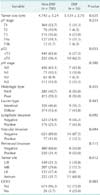
Table 2 shows the oncological characteristics of patients with and without DSF. The complication occurred more frequently in patients with tumors graded more than pT2 (P = 0.033). There was a significant difference in the incidence of DSF in patients with tumors located at the antrum compared with those with tumors located at the stomach angle to low body (P = 0.012). Patients who had a gastric outlet obstruction (GOO) before gastrectomy also had a higher occurrence rate (P = 0.003).
Surgical characteristics
The surgical characteristics of patients in the 2 groups are shown in Table 3. The surgical method, type of reconstruction, type of gastrectomy, extent of lymph node dissection, operation time, and intraoperative blood loss were compared. However, there were no statistically significant differences between the 2 groups.
Comorbidity factors
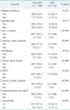
Univariate analysis was used to evaluate comorbidity factors in the 2 groups. The comorbidity factors used in our study were diabetes mellitus (DM), hypertension, liver cirrhosis, coronary artery disease, arrhythmia, chronic heart failure, chronic obstructive pulmonary disease, chronic kidney disease, and cerebrovascular accident. Patients under medication or observation after diagnosis with these comorbidities were all included in our analysis. Only DM showed a statistically significant difference between the two groups (P = 0.021). In addition, patients with multiple comorbidities had a significantly higher rate of DSF (P = 0.003). The results are summarized in Table 4.
Multivariate analysis of risk factors
Univariate analysis showed that age, ASA score, pathology T stage, tumor location, GOO, DM, and multiple comorbidities were significant factors for developing a DSF. In the multivariate analysis, multiple comorbidities (OR, 3.92; 95% CI, 1.30–11.80) and GOO (OR, 5.62; 95% CI, 1.45–21.71) were the risk factors for DSF (Table 5). Tumor location was excluded from this analysis because GOO only occurred in tumors located at the antrum.
Clinicopathology results
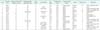
The clinicopathology results of patients who developed DSF are shown in Table 6. DSF occurred in 16 patients (2.2%) and there were 2 deaths in this subgroup. The mean interval of DSF after operation was 6.6 days. Both of the deaths were associated with complications from DSF [1]. One patient died from sepsis and bleeding and the other patient died from pneumonia. Three patients were submitted to a reoperation at postoperative days 1 or 2. Primary repair of the duodenal stump with choledochostomy and feeding jejunostomy was performed for these patients. Six patients were managed by ultrasound-guided PTBD. Seven other patients recovered with supportive care such as percutaneous drainage, TPN, and octreotide treatment. The mean duration of hospital stay after surgery was 29.4 days.
DISCUSSION
DSF after gastrectomy for gastric cancer is a rare complication with an incidence of 1.8%–3% [123]. However, once DSF occurs it is a life-threatening complication with a mortality rate of 7%–67% [123]. In our study, the incidence of DSF was 2.2% and the mortality rate was 12.5% in those patients. One patient died from sepsis and bleeding and the other patient died from pneumonia following DSF. These are common complications from DSF [1].
Various factors have been reported to cause DSF [4]. Orsenigo et al. [5] first reported the risk factors associated with postoperative DSF to be heart disease, liver cirrhosis, intraoperative blood loss (>300 mL), and the absence of manual reinforcement. We found here that age, ASA score, pathology T stage, tumor location, GOO, DM, and multiple comorbidities were significant factors for DSF in the univariate analysis. Multivariate analysis showed that GOO (OR, 5.62; 95% CI, 1.45–21.71) and multiple comorbidities (OR, 3.92; 95% CI, 1.30–11.80) were the main risk factors for DSF after gastrectomy for gastric cancer. GOO occurs more frequently when gastric cancer locates in lower body, i.e., antrum. GOO can also cause inflammation at surrounding duodenal wall tissues. For these reasons, we secure more distal margin because of the danger that cancer can involve at resection line, and eventually more devascularization is required. This is why we think GOO could be a risk factor of DSF shown in our study [47].
GOO and multiple comorbidities are both easily revealed during preoperative evaluation for DSF (Fig. 1). Patients can be categorized into high-, and low-risk groups. High-risk patients are those who have GOO or multiple comorbidities. Low-risk patients are those who have no symptoms of obstruction and no more than a single comorbidity. This means that we can anticipate the risk before the operation and prepare to make an appropriate decision when DSF is doubtful. Moreover, the surgeon can pay more attention during the operation to prevent DSF from occurring.
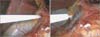
To prevent DSF during surgery, we used an absorbable reinforcement felt (Neoveil, Gunze Limited, Shenzhen, China) [8910] and a fibrin sealant (Tisseel, Baxter AG, Wien, Austria) [1112] on the duodenal stump. First, we applied the fibrin sealant onto the duodenal stump and then sealed it with the absorbable reinforcement felt. The fibrin sealant was then reapplied to prevent DSF (Fig. 2).
Many methods have been proposed for the treatment of DSF such as surgical management, a percutaneous approach, enteral and/or TPN, and therapy with somatostatin and its analogues. The usual surgical treatments are tube duodenostomy [13], repair with a rectus abdominis muscle flap [14], closure by Rouxen-Y duodenojejunostomy [15], and pancreaticoduodenectomy [16]. The usual methods of a percutaneous approach are abscess drainage, transhepatic biliary drainage [1718], fistula closure using cyanoacrylate or prolamine [19], percutaneous transhepatic biliary drainage (PTBD), occlusion balloons [20] or Foley balloon catheter [21].
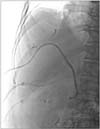
Three patients in our study underwent a reoperation involving primary repair of the duodenal stump using choledochostomy and feeding jejunostomy. Ultrasound-guided PTBD was performed for six of them (Fig. 3). The other seven patients recovered with supportive care. The mean duration of hospital stay after surgery was 29.4 days.
In conclusion, DSF is a life-threatening problem after gastrectomy for gastric cancer. Comorbidity and GOO were independent risk factors for DSF in our multivariate analysis. We generated a flow chart for risk factors based on these results. Surgeons could use this and pay more attention to prevent DSF in high-risk groups, and manage them more aggressively when DSF is anticipated.


 XML Download