Journal List > Ann Surg Treat Res > v.90(2) > 1093436

Jeong, Choi, Hwang, Yuk, Song, and Lee: Novel method of laparoendoscopic single-site and natural orifice specimen extraction for live donor nephrectomy: single-port laparoscopic donor nephrectomy and transvaginal graft extraction

Abstract

Laparoscopic live donor nephrectomy (DN) has been established as a useful alternative to the traditional open methods of procuring kidneys. To maximize the advantages of the laparoendoscopic single-site (LESS) method, we applied natural orifice specimen extraction to LESS-DN. A 46-year-old woman with no previous abdominal surgery history volunteered to donate her left kidney to her husband and underwent single-port laparoscopic DN with transvaginal extraction. The procedure was completed without intraoperative complications. The kidney functioned well immediately after transplantation, and the donor and recipient were respectively discharged 2 days and 2 weeks postoperatively. Single-port laparoscopic DN and transvaginal graft extraction is feasible and safe.

INTRODUCTION

Since 1995 when Ratner et al. [1] performed the first laparoscopic live donor nephrectomy (LLDN), the method has become established as a useful alternative to the traditional open methods of procuring kidneys for transplantation. LLDN has several advantages over open methods, including improved cosmesis, shorter hospital stay, reduced intraoperative blood loss, and faster overall postoperative recovery [2]. Furthermore, studies have shown that LLDN has positively impacted the number of kidneys available for transplantation. Most transplant centers harvest living donor kidneys via conventional laparoscopic surgical approach [3].

Laparoendoscopic single-site donor nephrectomy

Laparoendoscopic single-site donor nephrectomy (LESS-DN) is a relatively novel minimally invasive approach that allows the surgery to be performed via a single incision. This technique may be advantageous in decreasing surgical morbidity and improving cosmetic outcomes, thus possibly reducing the barriers to kidney donation [4].
Fan et al. [5] performed a meta-analysis of 27 studies comparing LESS and laparoscopic nephrectomy in patients undergoing radical or partial nephrectomy for cancer, donor nephrectomy, or mixed procedures. They found LESS nephrectomy to be a safe and effective alternative, with potential benefits of reduced pain, shorter recovery, and improved cosmesis. Recently, Aull et al. [6] performed a randomized, prospective study comparing LESS-DN and LLDN and reported that LESS-DN offers a safe alternative to traditional LLDN, with similar donor recovery and satisfaction. LESS-DN and LLDN appeared to be equivalent in terms of outcomes, donor complications, and recipient graft function.
To maximize the advantages of LESS-DN, we applied a natural orifice specimen extraction (NOSE) method. Our goal was to report several technical advancements in a case of single-port laparoscopic donor nephrectomy with transvaginal extraction of the kidney.

Patient and methods

A 46-year-old woman with no previous history of abdominal surgery volunteered for left kidney donation to her husband. On CT angiogram, the left kidney had one artery and vein. Her body mass index of 24.8 kg/m2 was in the normal range but close to overweight. The left kidney was chosen for donation according to our routine protocol.
This technical note did not require Institutional Review Board review or approval because retrospective reports of a single case do not meet the definition of research. Instead, our Ethics Committee advised us to obtain a specific informed consent from the patient. The procedure was fully explained to the donor and recipient, and possible complications related to transvaginal extraction including infection and pelvic organ injury were discussed in detail. The procedure was carried out by a multidisciplinary team consisting of a gynecologist, nephrologist, urologist, and transplant surgeons.

Results

Single-port laparoscopic donor nephrectomy with transvaginal extraction was completed without intraoperative complications. Total operative time was 280 minutes. Warm ischemic time was 2 minutes 28 seconds, and estimated blood loss was 50 mL. The harvested renal vein and artery were each 4 cm long. The kidney functioned well immediately after transplantation, without occurrence of delayed graft function.
Postoperative pain was 5/10 on postoperative day 1 and decreased to 2/10 on postoperative day 2. The donor subsequently had an unremarkable hospital course and was discharged on postoperative day 2. No 30-day complications were observed. The recipient was discharged in good condition after 2 weeks.

SURGICAL TECHNIQUE

On July 1, 2014, the patient underwent single-port left-sided donor nephrectomy with transvaginal extraction of the donor kidney. Broad-spectrum antibiotics were administered, and the vagina and perineum were prepped with Povidone solution. The patient was placed in a modified right flank position for laparoscopic nephrectomy with the legs fixed in stirrups. The table was tilted to the right in order to achieve an optimal view.
A 2.0-cm vertical transumbilical incision was made, and pneumoperitoneum was established using an open Hasson technique. A commercial single port (OCTO port; Dalim, Seoul, Korea) was introduced into the peritoneum. The abdomen was insufflated to a pressure of 12 mmHg. A 10-mm (30°) rigid laparoscope and conventional straight laparoscopic instruments were used. The other general procedures and sequences were the same as those used in conventional laparoscopic donor nephrectomy, as briefly described below.
  1. A lateral white line of Toldt was dissected, and descending colon was mobilized medially.

  2. The gonadal vein was dissected from the level of the iliac vessels up to the lower pole of the kidney, ligated, and divided near the renal hilum.

  3. During posterior dissection of the renal vein, the lumbar vein was dissected and divided.

  4. The adrenal vein was dissected and divided at the upper border of the left renal vein.

  5. After circumferential dissection of the renal vein, dissection was continued medially to the origin of the abdominal aorta.

  6. The renal artery was identified and dissected circumferentially.

  7. The ureter was dissected downward to beneath the level of the iliac vessels.

  8. The entire kidney was freely mobilized by dissecting the lateral and posterior aspects.

  9. A gynecologist performed a posterior colpotomy via intraabdominal approach.

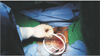
  10. After introducing a medium-sized wound retractor via a vaginal incision, a large wound retractor was inserted through the medium one and overlapped (Fig. 1).

  11. The surgical glove was fixed to the outer ring of the large wound retractor to maintain pneumoperitoneum.

  12. After making a small incision in the fingertip portion of the glove, a 15-mm trocar was inserted.

  13. The distal side of the ureter was ligated using a 5-mm Hemolock (Hemolock Ligating Clips, Teleflex, Morrisville, NC, USA). Then, the ureter was resected with an endoshear, and urine outflow was checked.

  14. A Lap-bag (Sejong Medical Co., Paju, Korea) was introduced transvaginally, and the kidney including the ureter was placed in the Lap-bag.

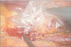
  15. The renal artery was ligated close to the aorta with a Multifire Endo-TA stapler (Auto Suture 30 mm–2.5 mm; Covidien, Mansfield, MA, USA) and cut distally with an endoshear.

  16. Similarly, the renal vein was also ligated and divided as close to the inferior vena cava as possible with the Multifire Endo-TA stapler (Auto Suture 30–2.5 mm, Covidien) and endoshear (Fig. 2).

  17. The Lap-bag orifice was closed, and the kidney was delivered transvaginally.

  18. The assistant removed the kidney from the bag, avoiding contamination from the external side of the bag.

  19. The colpotomy was repaired transvaginally with interrupted full-thickness reabsorbable sutures and checked at the intraperitoneal site with the laparoscope.

  20. Intra-abdominal conditions including hemostasis were checked laparoscopically, and the descending colon was reperitonealized with a V-loc wound closure device (Covidien).

DISCUSSION

The surgical technique for live donor nephrectomy has evolved from open surgery to LESS. Raman et al. [7] found no difference between LESS and standard laparoscopic nephrectomy in a retrospective case-control comparison using standard measures of in-hospital morbidity. In another retrospective review comparing LESS donor nephrectomy to laparoscopic approach, the LESS group had a significantly longer operative time (206 minutes vs. 182 minutes), but less blood loss (62 mL vs. 86 mL) and a shorter warm ischemic time (4.4 minutes vs. 5 minutes). Analgesic requirements were similar in the 2 groups [4]. Similarly, Barth et al. [8] conducted a postprocedural survey and found that LESS was associated with better scores for cosmetic outcomes and satisfaction with the overall process. Interestingly, blood loss was lower in the LESS group, along with reduced pain and fewer positional limitations under certain circumstances. In a randomized, prospective study, Aull et al. [6] reported that LESS donor nephrectomy can be a safe alternative to conventional laparoscopic donor nephrectomy in terms of intra- and postoperative complications.
In order to maximize the advantages of LESS, we recommended transvaginal extraction to the patient, which was completed without complications. Recently several authors have reported the feasibility and safety of transvaginal extraction [910]. The first laparoscopic living donor nephrectomy with vaginal extraction was performed in March 2009 by Allaf et al. [9]. In these initial reports, grand multipara donors were selected intentionally because laxity of the pelvic floor and vaginal wall is an important condition of graft passage. Our donor had also had three vaginal deliveries in the past. Not all women are good candidates for vaginal extraction, including those with prior pelvic surgery or younger women of childbearing age. Multiparous women may be better candidates for the procedure because of the laxity of the pelvic floor and vaginal walls [10].
We used advanced techniques to achieve maximum safety of the donor and graft. Before transection of the renal vessels, prepacking of the kidney inside the Lap-bag and advance preparation of the colpotomy allowed a quick delivery of the graft, keeping the warm ischemic time to a minimum, comparable to that of the standard laparoscopic extraction technique. During transection of the renal vessels, lateral traction of the Lap-bag offered good exposure of the pedicle, avoiding vessel injury. During the extraction process, we added several steps to decrease mechanical damage. We applied sterile lubricant gel around the wound retractor for smooth passage of the Lapbag and graft and checked the passage of three fingers (about 5–6 cm) in the opening to evaluate its size and the laxity of the vaginal wall. Because extraction under direct vision is important to graft safety, a surgical glove and 15-mm trocar were attached to the wound retractor to maintain pneumoperitoneum.
Kishore et al. [10] reported their initial 30 cases of laparoscopic donor nephrectomy with transvaginal extraction. They encountered one case of wound infection in a recipient in the transvaginal group. To prevent infection in the vaginal wall, we added several precautionary steps. Broad-spectrum antibiotics were administered, and the vagina and perineum were prepped with Povidone solution. We also developed a "dual wound retractor". After the posterior colpotomy was created, we placed a medium-sized wound retractor transvaginally. By overlapping a large wound retractor through the medium-sized retractor, we were able to obtain an aseptic pathway from the peritoneal cavity. To avoid contamination, the assistant removed the kidney from the bag without contamination from the external side of the bag.
Traditionally, the Endo GIA stapler, which has been shown safe and effective in numerous studies, has been used for vascular control. It works by placing six rows of staples, three proximally and three distally, with a blade that cuts between the staple lines. Hence, it leaves three rows of staples on the graft that must be trimmed prior to transplantation, which sacrifices several millimeters of length on the vein. By using a linear noncutting stapler (Auto Suture 30 mm–2.5 mm; Covidien) instead, we were able to preserve these several millimeters of length on the vein and extract the graft through a minimal incision.
For vein transection, the Multi Endo-TA stapler was placed on the renal vein as close to the vena cava as possible. After applying the Multi Endo-TA stapler, the vein was cut distally with laparoscopic scissors, which allowed blood to flow out of the graft through the renal vein and graft size to decrease. With this maneuver, we could extract the graft via minimal incision.
In our case, only a 2-cm abdominal incision was needed to complete the operation, and the scar was hidden inside the umbilicus (Fig. 3). No extended skin incision or fasciotomy was needed for graft extraction.
Kishore et al. [10] reported that the mean operative time of transvaginal extraction was 155±31 minutes and that of transabdominal extraction was 149±24 minutes. In our case, total operative time was longer by 130 minutes (total operative time, 280 minutes), which was mainly due to the NOSE maneuver. It took one hour to create a colpotomy, and 30 minutes to repair it.
A meta-analysis comparing LESS with conventional laparoscopic nephrectomy showed that LESS cases had a longer operative time and conversion rate, but had decreased pain and lower analgesic requirements, as well as shorter hospital stays, shorter recovery, and better cosmetic outcomes. The warm ischemic time and postoperative function of the donated kidney appeared roughly equivalent between groups [4]. Considering these advantages and our experience, single-port laparoscopic donor nephrectomy and transvaginal specimen extraction can be a novel technique for procuring kidneys.

CONCLUSION

By applying advanced techniques to LESS-DN, cosmesis and safety can be improved. Single-port laparoscopic donor nephrectomy and transvaginal graft extraction is feasible and safe. In the future, this maneuver may be an alternative standard in selected women.

Figures and Tables

Fig. 1

Dual wound retractor.

astr-90-111-g001
Fig. 2

Renal vein resection.

astr-90-111-g002
Fig. 3

Postoperative scar.

astr-90-111-g003

Notes

CONFLICTS OF INTEREST No potential conflict of interest relevant to this article was reported.

References

1. Ratner LE, Ciseck LJ, Moore RG, Cigarroa FG, Kaufman HS, Kavoussi LR. Laparoscopic live donor nephrectomy. Transplantation. 1995; 60:1047–1049.
2. Greco F, Hoda MR, Alcaraz A, Bachmann A, Hakenberg OW, Fornara P. Laparoscopic living-donor nephrectomy: analysis of the existing literature. Eur Urol. 2010; 58:498–509.
3. Wright AD, Will TA, Holt DR, Turk TM, Perry KT. Laparoscopic living donor nephrectomy: a look at current trends and practice patterns at major transplant centers across the United States. J Urol. 2008; 179:1488–1492.
4. Stamatakis L, Mercado MA, Choi JM, Sanchez EJ, Gaber AO, Knight RJ, et al. Comparison of laparoendoscopic single site (LESS) and conventional laparoscopic donor nephrectomy at a single institution. BJU Int. 2013; 112:198–206.
5. Fan X, Lin T, Xu K, Yin Z, Huang H, Dong W, et al. Laparoendoscopic single-site nephrectomy compared with conventional laparoscopic nephrectomy: a systematic review and meta-analysis of comparative studies. Eur Urol. 2012; 62:601–612.
6. Aull MJ, Afaneh C, Charlton M, Serur D, Douglas M, Christos PJ, et al. A randomized, prospective, parallel group study of laparoscopic versus laparoendoscopic single site donor nephrectomy for kidney donation. Am J Transplant. 2014; 14:1630–1637.
7. Raman JD, Bagrodia A, Cadeddu JA. Single-incision, umbilical laparoscopic versus conventional laparoscopic nephrectomy: a comparison of perioperative outcomes and short-term measures of convalescence. Eur Urol. 2009; 55:1198–1204.
8. Barth RN, Phelan MW, Goldschen L, Munivenkatappa RB, Jacobs SC, Bartlett ST, et al. Single-port donor nephrectomy provides improved patient satisfaction and equivalent outcomes. Ann Surg. 2013; 257:527–533.
9. Allaf ME, Singer A, Shen W, Green I, Womer K, Segev DL, et al. Laparoscopic live donor nephrectomy with vaginal extraction: initial report. Am J Transplant. 2010; 10:1473–1477.
10. Kishore TA, Shetty A, Balan T, John MM, Iqbal M, Jose J, et al. Laparoscopic donor nephrectomy with transvaginal extraction: initial experience of 30 cases. J Endourol. 2013; 27:1361–1365.
TOOLS
Similar articles