

 PDF
PDF ePub
ePub Citation
 Print
Print


INTRODUCTION
Combined hepatocellular cholangiocarcinoma (ChC) is a rare subtype of primary liver cancer. The incidence of ChC accounts for 1.6% to 6.5% of surgically resected primary liver cancers [1,2,3,4,5,6]. Patients with ChC typically have a shorter overall survival time and experience earlier recurrence after surgical resection than patients with hepatocellular carcinoma (HCC) [2,3,7]. However, some authors have reported similar survival outcomes between patients with ChC and those with HCC [8].
The dysplastic nodules have been introduced the precancerous lesions of HCC from proposal of the International Working Party in 1995. And cell type feature, tumor structures and cytological variants has been diagnostic basis of HCC in World Health Organization (WHO) classification [9]. In recently, some reports suggested that hepatic progenitors/stem cell play a role of HCC origin [9,10,11,12,13,14]. And the presence of cancer stem cells is associated with carcinogenesis, vascular invasion, and metastasis in primary liver cancer [10,11,12,15,16,17]. Moreover, the 2010 WHO classification suggested that ChC should be divided into 2 subtypes: the classical type and subtypes with stem cell features [18].
To date there have been no studies comparing the expression patterns of cancer stem cell markers between ChC and HCC or evaluating the expression of each cancer stem cell marker as a prognostic variable in ChC. In our study, we investigated the expression patterns of cancer stem cell markers between ChC and matched cases of HCC, and we evaluated whether patient prognosis had any correlation to the expression of each cancer stem cell marker in ChC.
METHODS
Patient selection and collection of clinical data
From January 2000 to June 2013, 443 patients with HCC underwent hepatic resections in our hospital. During the same period, 14 patients who underwent curative hepatic resection were pathologically diagnosed with ChC at our institution. One patient was excluded because of the loss of a pathology slide; therefore, 13 patients with ChC were enrolled in our study. We identified 13 patients with HCC matched for the following criteria: age, sex, a result of hepatitis viral serologic test, Child-Turcotte-Pugh (CTP) classification, tumor size, multiplicity, extent of hepatectomy, macrovascular invasion, and a-FP. A total of 26 patients were enrolled in our study. We retrospectively collected all clinical medical records including personal medical history, laboratory data, operative findings, and pathological reports. The personal medical history recorded patients' age, sex, and hepatitis history. The laboratory data consisted of hepatitis viral serologic results, including preoperative PT (international normalized ratio, INR), serum albumin level, indocyanine green ratio after 15 minutes (ICG R15), α-FP level, and CA19-9 level. The operative findings indicated the extent of hepatectomy, where major resection means resection of more than 3 segments and minor resection means resection of less than 3 segments, and complications. Pathological reports revealed tumor size (cm), multiplicity, liver cirrhosis, and macrovascular invasion, which means that the tumor has invaded the main portal vein or hepatic vein.
Immunohistochemical staining
All pathological specimens were formalin-fixed and paraffin-embedded. An experienced pathologist evaluated the pathology slides of 26 patients according to the 2010 WHO classification [18] which as mentioned included an important change in classifying ChC into either the classical type or subtype with stem cell features. To discriminate between the 2 types in our study, immunohistochemical staining was conducted with an autostainer (LVAUT4802SD, Lab Vision Autostainer 480, Thermo Scientific Inc., Waltham, MA, USA) according to the manufacturer's instructions. Immunohistochemical staining was performed on all tissue samples by using monoclonal antibodies against biliary markers cytokeratin (CK)7 (M7018, 1:100 dilution; Dako, Carpinteria, CA, USA) and CK19 (M0888, 1:100 dilution; Dako) and the following cancer stem cell markers: cluster of differentiation (CD) 117 (c-kit, A4502, 1:50 dilution; Dako), CD44 (orb69034, 1:500 dilution; Biorbyt, Cambridge, UK), CD133 (orb99113, 1:500 dilution; Biorbyt), and epithelial cell adhesion molecule (EpCAM, orb10618, 1:400 dilution; Biorbyt). The immunohistochemical staining was visualized by using horseradish peroxidase conjugates. The positive expression of each marker was defined as more than 50% staining in the whole hepatocelluar component of the specimen. The magnifying power for CK7 and CK19 was ×100 and that of other cancer stem cells (CSCs) markers was ×400.
Statistics
To compare the clinical data including the expression patterns of cancer stem cell markers between ChC and HCC, categorical variables were analyzed by using Pearson chi-square test and Fisher exact test, and continuous variables by using the Mann-Whitney test. To identify the prognostic variables in terms of overall survival and recurrence-free survival, univariate analyses were performed with the Kaplan-Meier survival curve using a 2-sided log-rank test. Statistical analysis was performed with SPSS ver. 14.0 (SPSS Inc., Chicago, IL, USA). A P-value of less than 0.05 was considered statistically significant. The study protocol was approved by the Institutional Review Board of the Korea Cancer Center Hospital.
RESULTS
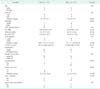
The clinical characteristics of patients with ChC and HCC are summarized in Table 1. For the patients with ChC, 9 patients (69.2%) were men and their median age was 52.0 years. The HBsAg was present in 6 patients (46.1%) and the CTP classification of all patients was A. In the preoperative laboratory data, the median value of PT (INR) was 1.09 and that of albumin was 4.2 g/mL. The median value of ICG R15 was 13.0%; the median values of tumor markers a-FP and CA 19-9 were 100.7 ng/mL and 14.12 U/mL, respectively. In the operative findings, 10 patients (76.9%) underwent major hepatectomy. Six patients (46.1%) had complications, such as atelectasis, pneumonia, urinary infection or wound infection. There was no hospital mortality among the study groups. In the pathology reports, 3 patients (23.1%) had multiple tumors and the median tumor size was 6.3 cm. Two patients (15.4%) showed liver cirrhosis and 1 patient (7.7%) showed invasion of the portal vein. Between ChC and HCC, the clinical characteristics showed no statistically significant difference because of the matching methods used.
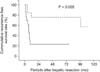
The median follow-up period was 25.0 months, ranging from 2.0 to 129.0 months. Hospital mortality did not occur after surgical resection. Two patients with ChC died during the follow-up period and none of HCC patients died. Ten patients with ChC and 4 patients with HCC had recurrences. We tried to analyze the prognostic variables for overall survival for all patients, but we could not gather meaningful results because of low event cases of death during the follow-up period. Instead, we analyzed the prognostic variables for recurrence-free survival. The mean recurrence-free survival time of ChC was 21.7 months, but that of HCC was 82.8 months (P = 0.005) (Table 2, Fig. 1).
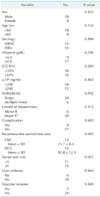
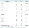
There were 5 cases of ChC with stem cell features (38.5%) and 4 cases of HCC with stem cell features (30.8%) (Fig. 2A-C). The expression patterns of cancer stem cell markers were not significantly different between ChC and HCC (P > 0.999). CK7 expression was positive in 1 case of ChC (7.7%) and in 3 cases of HCC (23.1%) (Fig. 2A). CD44 was expressed in 3 cases of ChC (23.1%) and in no cases of HCC (Fig. 2B). However, neither CK7 nor CD44 expression in ChC showed a significant difference compared to that expressed in HCC (P = 0.593 and P = 0.220 respectively) (Table 3). Likewise, there was no statistically significant difference in the expression of other cancer stem cell markers between ChC and HCC (all P > 0.999) (Table 3).
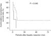
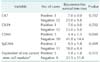
To identify the relationship between patient prognosis and cancer stem cell marker expression in ChC, we analyzed recurrence-free survival in 13 cases. The ChC subtype with stem cell features was not associated with a poorer prognosis than the classical type of ChC (P = 0.515). However, recurrence-free survival analysis of the expression of each cancer stem cell marker revealed that the CD44 positive group showed earlier recurrence than the CD44 negative group (P = 0.040) (Fig. 3). The recurrence-free survival time of the CD44 positive group was 4.4 ± 1.3 (mean ± standard deviation) months, whereas that of the CD44 negative group was 41.8 ± 14.4 months. There was no significant difference between the groups that were positive for other markers and the groups that were negative (Table 4).
In HCC, the overall expression of cancer stem cell markers was not associated with recurrence (P = 0.526), and the expression of individual cancer stem cell markers was not associated with recurrence (Table 5).
DISCUSSION
Most studies of ChC have been those comparing the disease to HCC or cholangiocarcinoma [2,3,4,7,8]. Some of these studies have shown that ChC has a poorer prognosis than HCC [2,3,7]. However, compared to cholangiocarcinoma, the prognosis of ChC is controversial [2,3,4,7,8]. Koh et al. [3] reported that ChC showed higher prevalence of portal or hepatic vein invasion, microvascular emboli and multiple tumors than HCC (P = 0.025, 0.003, and lower than 0.001, respectively). And Lee et al. [7] reported that ChC is associated with more advanced stages of cancer and more frequent cirrhotic changes of the liver than HCC (P = 0.009 and lower than 0.001, respectively); however, their analysis of those variables could have accounted for the poorer prognosis of ChC compared to HCC. Considering the lower incidence of ChC, a matching study would be more effective for comparing prognoses between ChC and HCC. In our study, we tried to match patients with ChC and HCC for age, sex, hepatitis viral serologic results, CTP classification, tumor size, multiplicity, extent of hepatectomy, macrovascular invasion, and α-FP. These variables could have a similar impact on the prognosis of both combined HCC and HCC. As previous studies have reported, our results showed that patients with combined HCC had a shorter recurrence-free survival time than those with HCC. In addition, ChC was the only prognostic variable for recurrence-free survival that we observed, unlike that reported by previous studies. Therefore, we could conclude that ChC itself has a poorer prognosis compared to HCC.
The presence of cancer stem cells is associated with carcinogenesis, vascular invasion, and metastasis in primary liver cancer [10,11,12,15,16,17]. In addition, the expression of cancer stem cell markers in HCC has been reported to be associated with poor prognostic variables such as poor differentiation, major vascular invasion, advanced cancer stage, early recurrence, and low survival [11,12,14,16,17,19]. Moreover, the 2010 WHO classification suggested that ChC should be divided into 2 subtypes: the classical type and subtypes with stem cell features [18]. The classification recommended that if phenotypical or immunophenotypical features of stem/progenitor cells were predominant, ChC with stem cell features should be considered. Yu et al. [5] reported that 8 of 14 cases of ChC (57.1%) showed simultaneous expression of cancer stem cell markers c-kit, CD90, CD133, and CK19. Their expression scoring system used the rate of positive cells and the intensity of staining. Positive expression was defined as a final score of more than 4 where the grade of positive cells rate was multiplied by the staining intensity. Ikeda et al. [20] reported that the subtype of ChC with stem cell features accounted for 24 of 36 cases (66.6%). They defined the subtype with stem cell features as having more than 5% stem cell marker expression. In our study, 5 of 13 cases (38.5%) were the subtype of ChC with stem cell features. We defined this subtype as having more than 50% expression of each cancer stem cell marker. In studies to date, the proportion of ChC cases with stem cell features has ranged from 38.5% to 66.6% and the definitions of this subtype in each study were different [5,10,20,21,22]. To clarify the prevalence and prognosis of patients with the stem cell feature subtype, an exact definition for this subtype is needed, specifically which cancer stem cell markers to use and how to measure their expression. In addition, previous studies did not cite whether the expression area analyzed was in the HCC component or the cholangiocarcinoma component of the tumor. In our study, we analyzed the expression of cancer stem cell markers in the HCC component, not the cholangiocarcinoma component. Akiba et al. [21] identified a slightly different expression pattern of stem cell markers in the HCC component as compared to the cholangiocarcinoma component and suggested that ChC has a broad histologic spectrum. Therefore, the exact definition for the subtype with stem cell features should specify which component of the tumor has been analyzed.
The expression of cancer stem cell markers in HCC have been reported that it is associated with tumorogenesis, tumor invasion, chemoresistance and poor prognosis [12,14,17]. However, we showed that the expression of cancer stem cell markers in HCC did not show a different pattern to that in ChC. In addition, the expression of cancer stem cell markers was not significantly associated with recurrence. However, there was a trend for the difference of cancer stem cell expression in some CSC marker like CD44, between ChC and HCC. So we carefully expected that the expression pattern could be showed a significant difference if sufficient cases were included in study. And we also could expect that in a large-scaled study of HCC, the prevalence of HCC with stem cell features maybe showed different patterns and associated with patient prognosis.
CD44 is a crucial receptor for binding hyaluronan [23]. CD44 expression is associated with the Wnt signaling pathway in the intestinal epithelium and is regulated by microRNAs such as microRNA-21 and microRNA-373-520c [24]. CD44 is also associated with the process of epithelial mesenchymal transition adhesion, adhesion to the adjacent tissues, metastasis and chemoresistance in solid tumors [24]. And the cooperation between CD44 and receptor tyrosine kinases induced antiapoptosis [24]. In a clinical study Endo and Terada [19] reported that CD44 expression was correlated with high histologic grades, vascular invasion, and poorer survival outcomes in patients with HCC. Moreover Henry et al. [25] reported that microRNA 199a-3p expression was reduced in 7 hepatocellular cell lines and that CD44 was essential for the c-Met signaling pathway, which is known to be a target of microRNA 199a-3p; they also suggested that CD44 may be an effective target treatment for CD44 positive HCC cells. In our study, not all cancer stem cell markers were associated with patient prognosis in ChC. Only the CD44 positive group in the ChC cases showed earlier recurrence than the CD44 negative group. We firstly reported that CD44 is associated with early recurrence in ChC. Even though the expressions of other cancer stem cell markers were not associated with prognosis, a specific marker, CD44 in our study, can be considered as a possible prognostic indicator. Further studies are needed to determine whether cancer stem cell markers can be used as prognostic factors.
ChC is a rare form of primary liver cancer [1,2,3,4,5,6]. The enrolled cases in other most studies were less than 20 cases. There were only 13 patients enrolled in our study. The limitation of our study was based on the insufficient number of cases of ChC. In addition, we were unable to analyze overall patient survival because of shorter follow-up periods. Nevertheless, our study is the first comparative analysis of the expression of cancer stem cell markers between ChC and matched HCC and for CD44 expression as a prognostic variable in ChC.
In conclusion, patients with ChC showed a poorer prognosis than those with HCC even though the prognostic variables in our study matched those previously reported. The expression of cancer stem cell markers in ChC did not show a significantly different pattern compared to that found in HCC. Finally, CD44 expression in ChC was an indicator of poor prognosis and of early recurrence.

 XML Download