

 PDF
PDF ePub
ePub Citation
 Print
Print


INTRODUCTION
Stomach cancer is one of the most common malignancies in Korea. The incidence of early detection of gastric cancer has increased with the increased frequency of medical checkups. Consequently, there is an interest in the quality of life following the detection and the cancer treatment options [12].
Following research on less invasive treatments, both the indications and techniques of laparoscopic gastrectomy have been refined [345]. Laparoscopic gastrectomy is becoming increasingly popular due to its advantages, which include faster recovery, reduced pain, better quality of life, and better cosmetic appearance, compared to open gastrectomy [46789]. Laparoscopic gastrectomy generally requires a small incision for anastomosis and there have been many attempts to reduce the size of this incision, with many surgeons striving to perform totally laparoscopic gastrectomies [10]. Anastomoses in laparoscopic gastrectomies are still an issue and extracorporeal anastomoses are preferred because of the difficulties of intracorporeal anastomoses [2811121314].
After laparoscopic surgery was introduced, continued refinements of the surgical tools and skills have made the surgery even less invasive [6]. Kanaya et al. [3] reported on an intraabdominal delta-shaped gastroduodenostomy using endoscopic linear staples with a smaller incision. Kim et al. [12] analyzed and reported the surgical results from the same procedure as above.
We attempted to perform modified intracorporeal gastroduodenostomy from a delta-shaped anastomosis, which overlaps the remnant stomach and the duodenal stump. We report the surgical results and clinical utility of the overlap method.
METHODS
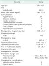
Between December 2011 and March 2013, 42 patients underwent totally laparoscopic distal gastrectomy (TLDG) with the overlap method for early gastric cancer. We reviewed their medical records and analyzed the data retrospectively (Table 1).
Procedures
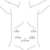
Under general anesthesia, the patient was placed in a supine and reverse Trendelenburg position. The operator and the camera assistant were on the right side of the patient and the assistant was on the left. Pneumoperitoneum was established with CO2 at a pressure of 12-13 mmHg. Five ports were placed on the abdomen including a port below the umbilicus (5 or 11 mm) for the camera (Fig. 1). In some surgeries, an additional port (5 mm) was placed at the epigastrium for liver traction.
Duodenal division and gastric resection
After dissection around the pylorus and duodenum, duodenal division was performed. The surgeon and the assistant grasped the anterior walls of duodenum and stomach, respectively. Then, pulling the duodenum and stomach in a caudal direction, duodenal division was performed with a 60-mm endoscopic linear stapler (Endo-GIA, Covidien, Mansfield, MA, USA), rotated 90° relative to a conventional division. Next, the lymph nodes were dissected, and trimming around the stomach was performed. Then, gastric resection was performed with a couple of endoscopic linear staples. The resected stomach that was surrounded with a vinyl pouch was removed through an extended lower left incision after anastomosis.
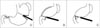
Intracorporeal gastroduodenostomy
A small gastrostomy and a duodenotomy are necessary to introduce the linear stapler during the gastroduodenostomy. The gastrostomy was located at the greater curvature, 6 cm from the previous staple line of the gastric resection. The duodenotomy was made at the anterior pole of the staple line of the duodenal division (Fig. 2). A 60-mm linear stapler (Tan-Cartridge Endo-GIA, Covidien) was inserted into two incisions in the stomach and duodenum, and an anastomosis was made between the greater curvature of the stomach and the anterior wall of the duodenum. The stapler was then advanced through an 11-mm port at the lower left region of the abdomen for easy anastomosis. After verifying that there was no bleeding at the anastomotic staple line, the remaining opening was closed with a 60-mm purple-cartridge Endo-GIA. To prevent anastomotic stenosis, the closure was performed using widened staple lines, in a V-shape (Fig. 3) (Supplementary material).
RESULTS
Forty-two patients (26 males and 16 females) underwent an intracorporeal gastroduodenostomy with the overlap method. The mean age was 58.3 ± 11 years and the mean body mass index was 24.2 ± 3.0 kg/m2. Preoperative staging by computed tomography (CT) and endoscopy showed that 39 patients had stage IA and 3 had stage IB gastric cancer. The mean operation time was 228.3 ± 42.5 minutes and the mean anastomosis time was 12.18 ± 2.3 minutes (Table 1).
Lymph node dissection was performed more than D1+ [15]. The mean number of linear staples used in the operations was 5.5 ± 0.7. After the operation, 34 patients had stage 1A, 5 had stage 1B, and 3 had stage II gastric cancer. The mean length of the free proximal margin was 5.8 ± 2.8 cm (range, 2-14 cm), and the mean length of the free distal margin was 4.9 ± 2.1 cm (range, 1.5-11 cm) (Table 1). There were no open conversion cases and no postoperative mortalities.
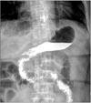
Upper gastrointestinal series (gastrografin) 5 days after the operations showed no anastomotic leakage or stricture and no delayed passage of contrast media (Fig. 4).
Abscesses were found in three patients on CT during the postoperative period. These patients also had a fever with a body temperature over 38℃ and constant leucocystosis. One of these patients underwent surgery for a suspected anastomosis leak, which was found to be fluid collection. The condition of the other two patients improved with conservative treatment. Five patients complained of gastric stasis symptoms during the postoperative period, which improved with fasting and diet control (Table 1).
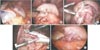
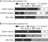
Patients underwent endoscopy at 6 and 18 months after their operation. The residue, gastritis, bile classification was used to evaluate the endoscopic findings (Fig. 5) [16].
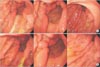
For all patients, anastomosis areas were wide enough, with no occurrences of stenosis or fibrotic stenosis due to ischemia in the duodenum (Fig. 6).
DISCUSSION
In laparoscopic gastrectomy, laparoscopy assisted distal gastrectomy (LADG) is currently preferred to TLDG [17]. Although TLDG is being used presently, it is challenging due to the difficulty of performing an intracorporeal anastomosis [10].
LADG is usually performed with a circular stapler or handsewing through a mini-laparotomy. In TLDG, an endoscopic linear stapler is used to perform the anastomosis, which is easily accomplished through a port in the body instead of a larger incision [10].
Kanaya et al. [3] introduced the technique of using an endoscopic linear stapler for intracorporeal gastroduodenostomy. This technique was named the delta-shaped anastomosis due to the shape of the gastroduodenostomy after anastomosis.
The delta-shaped anastomosis is an effective method for TLDG. However, there are some difficulties with this procedure. In the delta-shaped gastroduodenostomy, the posterior wall of the stomach and the posterior wall of the duodenum are anastomosed. For tension-free anastomosis, the surgeon must create sufficient space in duodenum [14]. However, this could increase the risk of impaired circulation, which can result in ischemia at the anastomosis. Because the anastomosis alignment lies over the superior side of duodenum, vessels of the duodenum are at risk for ligation and ischemia [18]. Creating sufficient space for the anastomosis is difficult, but insufficient space causes focal tension in the anastomosis, and mild bending after the anastomosis.
The intracorporeal gastroduodenostomy performed in this study is called the overlap method because the remnant stomach overlaps with the duodenal stump to form an anastomosis using an endoscopic linear stapler. This method is that duodenal division is performed in a 90° rotated state relative to a conventional division, so the anastomosis is on the anterior wall of the duodenum. This 90° rotation decreases the chance of vessel injury at the duodenum's superior border of the gastric and duodenal anastomosis. When an anastomosis is made on the anterior wall of the duodenum, sufficient space for insertion and manipulation of a linear stapler could be secured, that might make the anastomosis easy. This reduces the possibility of damage to the surrounding structure and duodenum relative to an anastomosis on the posterior wall of the duodenum. The risk of ischemia due to duodenal dissection is also reduced. After anastomosis on the duodenal anterior wall, closure of the common incision is performed just like the V-shape. This closure results in a triangular lumen at the site of the anastomosis and ensures sufficient luminal space to help prevent postoperative stenosis. Endoscopy 6 months and 18 months after the operation showed there was sufficient anastomosis area and no stenosis.
Our method of approaching the anterior wall of the duodenum provides an easier angle than the former method of approaching from the posterior wall. Using the overlap method, anastomosis alignment is parallel with the direction of the stomach and duodenum and the traction force from the stapler is in one direction. This arrangement maintains an even tension at the site of the anastomosis and provides an easy angle between the stomach and duodenum, so it could prevent kinking in anastomosis. A study using a similar method has also reported that this approach is considered to be an easy and relatively safe technique [10].
By reducing the extent of duodenal dissection, the overlap method can preserve sufficiently long first portion of the duodenum for anastomosis. According to the Japanese gastric cancer treatment guidelines 2010 (version 3), for T1 tumors, a gross resection margin of 2 cm should be obtained [15]. In this study, a proximal tumor-free margin met these guidelines (range, 2-14 cm). Three patients showed a proximal tumor-free margin of 2 cm and their postoperative T stage was T1.
Kanaya et al. [311] reported satisfactory results in 100 cases with a mean time to perform anastomosis of 13.0 ± 3.9 minutes. Park et al. [14] presented research on delta-shaped anastomoses in 57 patients between 2004 and 2009 and reported a mean procedure time of 20 minutes. Song et al. [10] reported a mean procedure time of 17 minutes. In this study, the mean time to perform an anastomosis was 12.18 ± 2.3 minutes. Despite initial experience, the overlap method's mean time in our study was shorter than or similar to the times reported by Song et al. [10] for delta- and linear-shaped anastomoses. These results may be attributed to the fact that the angle for anastomosis was easier to maintain. And it was required fewer staplers than delta-shaped anastomosis. More experienced operators have reported anastomosis procedure times significantly shorter [14].
The mean number of linear staples used in our operations was 5.5 ± 0.7. This is less than the mean of 6.7 reported by Kim et al. [12], and similar to the mean of 5.6 reported by Song et al. [10]. This shows that it is generally possible to complete an anastomosis with fewer staplers by using the overlap method.
In this study, 3 patients had intraabdominal fluid collection with no anastomosis leakage and all recovered after conservative treatment. Kim et al. [1] reported that there were 3 complications among the 25 cases of TLDG with Billroth-I anastomosis. One complication was anastomotic leakage, another was anastomotic stenosis, and the third was delayed gastric empting. A laparoscopic tube duodenostomy was performed for the anastomotic leakage, and endoscopic balloon dilatation was performed for the anastomotic stenosis, and conservative treatment was used for the delayed gastric empting. Kanaya et al. [11] reported 1 case of minor leakage out of 100 cases of the delta-shaped method that was treated with conservative treatment. Song et al. [10] reported 1 case of anastomotic stenosis among 25 cases of linear-shaped gastroduodenostomy. This patient underwent reoperation for gastrojejunostomy.
In conclusion, the overlap method of intracorporeal gastroduodenostomy, using an endoscopic linear stapler can be considered a feasible and safe method to achieve positive results. There are several merits such as evenly spacing the tension around the anastomosis, saving intestine length, and preventing the kinking of the anastomosis. However, a long-term comparative study is needed for an in-depth evaluation of this technique.
 XML Download