

 PDF
PDF ePub
ePub Citation
 Print
Print


INTRODUCTION
Randomized clinical trials have established that carotid endarterectomy (CEA) is a durable procedure for the prevention of recurrent neurological symptoms and stroke in patients with moderate to severe symptomatic and asymptomatic carotid artery stenosis [1234]. Patients with bilateral significant internal carotid artery (ICA) stenosis, either symptomatic or asymptomatic, may be at risk of cerebrovascular events or even cognitive impairment [56]. Despite the efficacy of CEA, there has been controversy over the operative risks of this procedure and the optimal interval between the primary and secondary operations for patients with bilateral ICA stenosis undergoing staged bilateral CEA [78]. Therefore, patients with bilateral ICA stenosis have been excluded from most prospective trials, and data on the safety and efficacy of bilateral revascularization procedures are currently scarce.
The aim of the present single-center study was to retrospectively compare the short-term operative risk and long-term outcomes of patients with bilateral significant ICA stenosis who underwent staged bilateral CEA within 30 days or less to those who underwent unilateral CEA. Our hypothesis was that the outcomes of patients who underwent staged bilateral CEA would not be inferior to those who underwent unilateral CEA.
METHODS
Study design and patient population
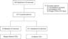
This retrospective observational study was based on data ob tained from medical records. The study protocol was approved by the Institutional Review Board of Asan Medical Center, and all patients provided written informed consent. Between September 2007 and August 2014, 645 consecutive patients with a significant carotid bifurcation stenosis (i.e., ≥70% in asymptomatic patients and ≥50% in symptomatic patients), as defined by criteria established by the North American Symptomatic Carotid Endarterectomy Trial (NASCET) [1], diagnosed by carotid duplex ultrasonography and magnetic resonance (MR) angiography, underwent a CEA. Patients with contralateral ICA occlusion (31 patients), contralateral carotid artery stenting (CAS) (8 patients), and contralateral CEA >30 days (32 patients) were excluded, and the remaining 574 patients were included in the study (Fig. 1). Of these 574 patients, patients diagnosed with bilateral ICA stenosis who underwent staged bilateral CEA within 30 days or less were included in the treatment group. To compare the short-term operative risk and long-term clinical outcomes, patients with unilateral ICA stenosis who underwent unilateral CEA were placed in the control group.
Demographics, risk factors and other data, including clinical characteristics and short-term operative risk, and long-term outcomes were prospectively recorded for all consecutive patients in an Excel database (Microsoft Corp., Redmond, WA, USA) and retrospectively analyzed.
CEA procedure and follow-up
The surgical procedure used has been previously detailed [9]. CEA was preferably performed under general anesthesia with endotracheal intubation, and regional cervical block (superficial and deep) was used in selected patients. In patients with general anesthesia, a Javid carotid shunt (Bard Inc., Murray Hill, NJ, USA) was routinely used. Endarterectomy with patch angioplasty was performed in the standard fashion with 3.5× optical power magnification and Prolene 5/0 and 6/0 continuous sutures. Tacking sutures to secure the distal intima and ICA plication were performed if needed. Postoperatively, all patients were given antiplatelet therapy in combination with stringent control of blood pressure and close observation in an intensive care unit for at least 24 hours. All patients were followed up both clinically and by MR imaging with angiography prior to discharge.
In patients with bilateral ICA stenosis, the side to be operated on first, defined as ipsilateral, was determined according to the following priority criteria: the presence of neurological symptoms, the degree of the stenosis, the presence of asymptomatic cerebral infarcts, and the dominant cerebral hemisphere [10]. The most symptomatic or higher-grade stenosed artery was identified as the primary lesion (also referred to as the ipsilateral lesion) and was treated first, and the less symptomatic or lower-grade stenosed artery was staged and scheduled for CEA within 30 days or less after the first CEA (referred to as the contralateral lesion).
Follow-up included carotid duplex ultrasonography to assess patency and exclude the development of new or contralateral lesions, as well as independent neurological examination using the National Institutes of Health Stroke Scale (NIHSS) [11] and the modified Rankin scale at 6 months, 12 months, and annually thereafter. Once stability had been established over 3 years, surveillance was performed at longer intervals of about 2 years.
Endpoint definitions
The primary endpoint was the composite of any stroke, myocardial infarction, or death during the periprocedural period or ipsilateral stroke within 3 years after the CEA. Postoperative stroke was defined as an acute neurological event with focal symptoms and signs, lasting for 24 hours or more, that were consistent with focal cerebral ischemia [12]. Strokes were categorized as ipsilateral or contralateral, as periprocedural (≤30 days) or late (≥31 days from the CEA), and as major or minor [7]. A major stroke was a stroke that was present after 7 days and increased the NIHSS of the patient by ≥4 points. A minor stroke was a stroke that resolved completely within 7 days or increased the NIHSS of the patient by ≤3 points. Myocardial infarction was defined by a cardiac troponin I level that was twice the upper limit of the normal range or higher according to our laboratory, in addition to either chest pain or symptoms consistent with ischemia or electrocardiography evidence of ischemia, including new ST segment depression or elevation or elevation of more than 1 mm in two or more contiguous leads according to the core laboratory [12].
Statistical analysis
Results are expressed as mean ± standard deviation for continuous variables and number and percentage for categorical variables. A Student t-test was used to compare the results of quantitative assays, and the chi-square test was used to compare categorical variables. Cumulative event risks were estimated from Kaplan-Meier survival curves and compared using the log-rank test. The Cox proportional hazards model was used to obtain hazard ratios and 95% confidence intervals for the outcomes. Statistical calculations were performed using PASW Statistics ver. 18.0 (SPSS Inc., Chicago, IL, USA), and differences were considered statistically significant at P < 0.05.
RESULTS
Of the 574 patients who underwent CEA, 43 patients (7.5%) with bilateral significant ICA stenosis who underwent staged bilateral CEA within 30 days or less were identified; 531 patients (92.5%) with unilateral ICA stenosis underwent unilateral CEA. The groups who underwent unilateral CEA and staged bilateral CEA did not differ significantly with regard to demographics, risk factors, or clinical characteristics, except that the staged bilateral CEA patients were more likely to be male (84.6% vs. 97.7%, P = 0.019) and to have a higher percent stenosis of the ipsilateral ICA (76.1 ± 9.6 vs. 79.1 ± 8.2, P = 0.054). In the staged bilateral CEA patients, the mean time interval between the primary and secondary CEA was 12 days (range, 5-29 days). There were no significant differences in the 30-day rates of cranial nerve palsy (2.6% vs. 4.7%, P = 0.340) and nonneurological CEA-related complications (5.8% vs. 9.3%, P = 0.322) between the two groups (Table 1).
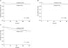
Patient age was the only risk factor significantly associated with ipsilateral stroke in univariate (P = 0.045) and multivariate (P = 0.024) analyses (Table 2). Staged bilateral CEA was not associated with ipsilateral stroke (P = 0.178) during postoperative follow-up. The groups who underwent unilateral CEA and staged bilateral CEA did not differ significantly in terms of primary endpoints during the periprocedural period (1.5% vs. 2.3%, P = 0.677) or in the estimated 3-year primary endpoints (2.8% vs. 4.7%, P = 0.456) (Table 3). There were no statistically significant differences between the unilateral CEA and staged bilateral CEA groups with regard to any solitary component of the primary endpoint. Kaplan-Meier survival analysis showed that the two groups had similar rates of ipsilateral stroke-free (P = 0.225), any stroke-free (P = 0.326), and overall (P = 0.739) survival (Fig. 2).
DISCUSSION
Although bilateral ICA stenosis is frequently encountered and substantially increases the risk of complications during and after unilateral CEA, controversy persists regarding the optimal treatment for this condition [7]. The aim of the present study was to evaluate the safety and efficacy of staged bilateral CEA in patients with bilateral significant ICA stenosis; we excluded the patients who underwent contralateral CEA >30 days because most of them presented with gradually aggravated ICA stenosis during the follow-up period after ipsilateral CEA. Our inclusion criteria was defined as staged bilateral CEA within 30 days or less based on the definition of periprocedural primary endpoint. Our present study findings indicate that the outcomes of potentially high-risk patients with bilateral significant ICA stenosis who underwent staged bilateral CEA within 30 days or less compare favorably with those of patients who underwent unilateral CEA.
In patients with bilateral significant ICA stenosis, the impaired cerebral hemodynamic status caused by chronic hypoperfusion may contribute to the development of cognitive dysfunction even if the patients are asymptomatic and the dysfunctions are not reflected in neuroimaging scans by brain structural changes [56]. Although the presence of a contralateral significant ICA stenosis has been associated with a high risk of adverse events after CEA or CAS, previous studies have shown that early selection of subjects deserving consideration for more aggressive revascularization procedures designed to improve cerebral hemodynamics would prevent cognitive dysfunction as well as recurrent neurological symptoms and stroke [56]. CEA is a durable procedure for the prevention of recurrent neurological symptoms and stroke in patients with significant ICA stenosis, and its durability has been established by randomized clinical trials, such as the NASCET and European Carotid Surgery Trial [12]. While the role of bilateral CEA simultaneously performed in one surgical procedure was controversial in patients with bilateral ICA stenosis [13], staged bilateral CEA using a short interval between the primary and secondary procedures was shown to be a safe and effective treatment concept [1415]. Recently, CAS with embolic protection is increasingly regarded as a treatment alternative in patients with ICA stenosis at high surgical risk [16171819], and some authors recommend staged bilateral CAS for the management of bilateral ICA stenosis due to a reported higher rate of cranial nerve palsy after staged bilateral CEA [714]. Although there has been controversy over the operative risks of CEA in these patients, the clinical outcomes may not be as poor as was once believed. In our current series, no significant differences were found when the rates of cranial nerve palsy were compared between the groups with unilateral CEA and staged bilateral CEA. Furthermore, we did not find statistically significant differences in 30-day and 3-year clinical outcomes between these two groups. At this time, despite the recent technical advances in CAS, CEA remains the gold standard for the management of carotid artery disease, based on the latest available data [820], and staged bilateral CEA within 30 days or less may be the optimal treatment strategy for selected patients with bilateral ICA stenosis.
Several limitations of our analyses should be noted. First, this study was retrospective in its design and this did not allow for direct randomized comparisons of treatment outcomes with those of other possible therapeutic strategies such as CAS or best medical treatment. In addition, there was no adjustment for baseline differences between the two groups. Furthermore, CEA of the contralateral carotid lesion within 30 days or less after CEA of the primary target lesion was allowed in our present series. Thus, our analysis could not give any insights into the clinical outcomes of simultaneous bilateral CEA. Although staged procedures have disadvantages, including a delay in the definitive treatment of carotid lesions, higher costs, and patient inconvenience, the concerns about simultaneous CEA seem to focus mainly on the hemodynamic impairment from stimulation of the carotid sinus baroreceptor and the risk of bilateral cranial nerve palsy and cerebral hyperperfusion syndrome [21222324]. Finally, our current findings were obtained from a single-center study with a consequently small sample size. This necessarily limits the overall applicability of our results.
Despite its potential limitations however, our present study shows that staged bilateral CEA within 30 days or less is an effective treatment option for patients with bilateral significant ICA stenosis and does not yield inferior outcomes to unilateral CEA. Considering that the usefulness of bilateral CEA in many asymptomatic patients may be debatable due to the recently improved efficacy of the aggressive medical treatment, future prospective studies of larger cohorts are warranted to confirm the findings from the present study.


 XML Download