

 PDF
PDF ePub
ePub Citation
 Print
Print


INTRODUCTION
Mucinous gastric adenocarcinoma (MGC) is a rare type of cancer that is defined by the World Health Organization (WHO) as a gastric adenocarcinoma with >50% extracellular mucin pools within the tumors [1]. MGC comprises approximately 2%-5% of all gastric carcinomas [2345]. The clinicopathologic characteristics of MGC remain controversial. MGC has shown a poorer prognosis than nonmucinous gastric adenocarcinoma (NMGC) [678]. In contrast, another study suggested no difference in the survival rate of MGC and NMGC.
Clinically, it is possible to find cases with extracellular mucin pools lower than a half of the tumor area (Fig. 1). However, the exact prevalence rate is unknown. It could be missing from the pathologic report because gastric cancer with lower than 50% tumor volume of extracellular mucin pool adenocarcinoma (LEMPC) is not included in the diagnostic criteria of WHO classification. Moreover, if a pathologist is not aware of LEMPC, extracellular mucin pools may be overlooked. According to the WHO classification, those cases are classified as NMGC, not MGC. Treatment of LEMPC is similar to that of NMGC, even though LEMPC tends to be more progressive [9].
The clinicopathologic feature of LEMPC patients is not clear. To clarify the effect of extracellular mucin pools, we compared the clinicopathologic features of NMGC patients with MGC and LEMPC patients to determine the effect of extracellular mucin pools. We also evaluated the independent prognostic factors from the point of recurrence.
METHODS
Subjects
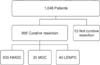
A retrospective review was done of 1,048 gastric cancer patients treated at Pusan National University Yangsan Hospital between December 2008 and December, 2013. Curative resection was performed in 995 patients (Fig. 2). MGC defined as extracellular mucin pools occupying over a half the tumor area was observed [1]. NMGC is classified as no extracellular mucin pool in tumor. LEMPC is defined as NMGC with including extracellular mucin pool.
All the diagnosis followed by the last report of pathologists. Clinicopathologic variables including gender, age, body mass index (BMI), tumor location, surgical methods, tumor size, depth of invasion, number of lymph node metastasis, number of resected lymph node, lymph node metastasis, Lauren classification, lymphatic invasion, vascular invasion, and perineural invasion were compared between MGC and NMGC patients, and between MGC and LEMPC patients. We investigated the variables in accordance with an established guideline [10]. Tumor staging was evaluated by using the TNM system of the International Union Against Cancer [10].
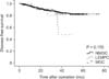
Prognostic factors associated with the recurrence of gastric cancers were also investigated. A disease-free survival (DFS) curve was prepared to identify the most significant prognostic factors for gastric cancer in our hospital based on recurrence. The diagnosis of recurrence was confirmed by radiological findings, computed tomography or positron emission tomography, and endoscopic biopsy with histopathological findings.
Statistical analyses
Data was statistically analyzed using the chi-square test and Student t-test. Odds ratios (ORs) were estimated with 95% confidence intervals (CIs). DFS curves were plotted using the Kaplan-Meier method and the log-rank test was used for univariate risk factors for recurrence. In multivariate analysis, the Cox proportional-hazards regression F model was used for independent risk factors of recurrence. All statistics were conducted using IBM SPSS Statistics ver. 21.0 (IBM Co., Armonk, NY, USA). Statistical significance was defined as P < 0.05.
RESULTS
All 995 patients underwent curative resection. MGC and LEMPC patients comprise 2% and 6%, respectively, of all patients.
Clinicopathological comparison between NMGC and other groups
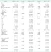
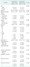
Mean ages of MGC, NMGC, and LEMPC patients were similar. Gender, BMI, and tumor location did not significantly differ in the three groups. MGC and LEMPC patients received more open gastrectomies with extended lymph node dissection than NMGC patients (both P < 0.001). MGC tumor size of was larger than NMGC tumors (P < 0.001). T stage, N stage, and node metastasis were higher in MGC patients than in NMGC patients (P < 0.001). LEMPC showed more advanced T stage (P < 0.001) and node metastasis than LMGC (P < 0.001). However, the N stage of LEMPC was not significantly different from that of NMGC (P < 0.0109). More positive perineural invasion was apparent in LEMPC than in NMGC (P < 0.010) (Table 1).
Clinicopathological comparison between NMGC and all extracellular mucin pool gastric adenocarcinomas
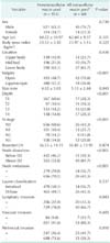
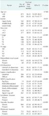
NMGC was compared with all extracellular mucin pool gastric adenocarcinomas. All extracellular mucin pool gastric adenocarcinomas involved more open gastrectomies (P < 0.001) with extended lymph node dissection (P < 0.001) than NMGC. All extracellular mucin pool gastric adenocarcinomas featured a larger tumor size and more positive node metastasis (P < 0.001), lymphatic invasion (P < 0.043), and perineural invasion (P < 0.001) than NMGC. All extracellular mucin pool gastric adenocarcinomas showed more advanced T stage (P < 0.001) and N stage (P < 0.001) (Table 2).
Clinicopathological comparison between LEMPC and MGC
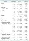
MGC featured higher T stage (P < 0.014) and more positive lymph node metastasis (P < 0.043) than LEMPC. Other clinicopathologic characteristics were not significantly different (Table 3).
Prognostic factors for DFS in patients with curative gastric cancer
In univariate analysis, prognostic factors of all patients were tumor lesion (P = 0.031), tumor size (P < 0.001), T stage (P < 0.001), N stage (P < 0.001), extent of lymph node dissection (P < 0.001), WHO classification (P < 0.001), lymphatic invasion (P < 0.001), vascular invasion (P < 0.001), and perineural invasion (P < 0.001). Extracellular mucin pool was not a prognostic factor of recurrence (Table 4).
DISCUSSION
Our results show that approximately 2% of the gastric cancer patients were MGC patients. The total gastric cancer patient's rate increased to 6% when all extracellular mucin pool gastric adenocarcinoma patients were included. These results did not show the difference that has been previously reported [9]. However, the prevalence of LEMPC is not well known. This is because LEMPC is not a WHO diagnostic criteria and it can be overlooked by endoscopists, clinicians, and even pathologists. If pathologists do not diagnosed with MGC, can be omit reported extracellular mucin pool composition.
The clinicopathologic features of MGC include large size [61112], deeper invasion [1314], and higher N stage than that of NMGC. In this study, MGC differed from NMGC in tumor size, T stage, N stage, and node metastasis. When the MGC is diagnosed, it is usually in a more advanced stage than NMGC. More extracelluar mucin pools within a tumor indicate a more progressive state of disease. Overexpressed transmembrane mucins are considered to be a signal of cell growth and survival. Extracellular mucin pools derived from overexpressed transmembrane mucin are relevant to the progression of carcinoma [15]. In this study, MGC featured more advanced T and N stages than NMGC.
LEMPC also showed similar results with MGC, having more advanced T stage and more positive node metastasis than NMGC. LEMPC and MGC patients received more radical surgery than NMGC patients. LEMPC and MGC showed similar cilinicopathologic features. Just, MGC had a larger tumor size, more advanced T stage and more positive node metastasis than LEMPC. This result suggests that LEMPC is an earlier state of MGC irrespective of other WHO classification. For this reason, it has been suggested to consider extracellular mucin pools carcinoma of more than 30% as MGC [9].
Additionally, all extracellular mucin pool carcinomas featured larger tumor size, more advanced T stage and N stage, more aggressive lymph node dissection with open surgery, more positive node metastasis, lymphatic invasion, and perineural invasion than NMGC. All extracellular mucin pool gastric adenocarcinomas were in a more advanced state than NMGC, regardless of Lauren classification. Therefore, we can consider all extracellular mucin pool gastric adenocarcinomas included with the diagnostic criteria of MGC. In this study, we analyzed the effect on the prognosis in terms of recurrence of gastric cancer. Univariate analysis showed that tumor size, T stage, N stage, range of lymph node dissection, Lauren classification, WHO classification, lymphatic invasion, vascular invasion, and perineural invasion were significantly correlated with gastric cancer recurrence in all patients treated by curative gastrectomy at our hospital. However, only T stage and N stage were identified as independent prognostic factors for gastric cancer recurrence in the multivariate analysis. It is well known that prognostic factors for gastric cancers are influenced by tumor invasion depth, lymph node metastasis, and complete tumor removal [161718]. Therefore, we assume extracellular mucin pools affect T stage and N stage, then the prognosis of gastric cancer recurrence
Limitations of the study include its retrospective design, and short follow-up. Specimens were not reassessed by pathologists again for the study. If all the specimens were reassessed by pathologists again for the study, then we can get high quality data. Moreover, we can calculate cutoff value of a linear/log relationship between mucin percent and clinical endpoints.
Despite these limitations, the results clearly show that the extracellular mucin pool gastric adenocarcinomas have more aggressive characteristics in T stage and N stage than NMGC. This suggests that in the treatment of these cancers, aggressive treatment is needed. More rigorous treatment is expected at the time of the operation rather than additional surgery if extracellular mucin pool gastric adenocarcinoma is predicted before surgery.
The amount of extracellular mucin pool content is not an independent prognostic factor for the recurrence rate. However, it needs to be seriously considered when diagnosed. When a preoperative biopsy result includes extracellular mucin pool carcinoma, we should be more careful in implementing the treatment, such as protection from mucin spillage while operating and more extensive lymph node dissection. Cautious consideration about treatment of extracellular mucin pool gastric adenocarcinoma patients may improve prognosis.
In conclusion, the clinicopathological characteristics of LEMPC patients are similar to MGC patients, except for T stage and positive lymph node metastasis. Patients with MGC had more progressed T stage and node metastasis than patients with LEMPC. We suggest that LEMPC can be thought of as a previous step of MGC. It is reasonable to consider LEMPC patients in the diagnostic criteria of MGC. To improve prognosis, we need more rigorous treatment for extracellular mucin pool gastric carcinoma. The results and conclusions of this paper are helpful for pathologists and surgeons when they make accurate diagnosis and adequate treatment.

 XML Download