

 PDF
PDF ePub
ePub Citation
 Print
Print


INTRODUCTION
Visceral artery aneurysms (VAAs) are rare forms of vascular pathology and most commonly involve the splenic artery [1]. Specifically, aneurysms of the pancreaticoduodenal artery are extremely rare, with a reported incidence of 2% among cases of VAA [2], and multiple VAA are rarer still [3]. Recent advancements in endovascular procedures have successfully reduced the morbidity and mortality of open surgery, thereby shifting the management paradigm [4]. However, open surgery should be considered when minimally invasive procedures are unlikely to be successful. Here, we report a unique case in which multiple VAA included the pancreaticoduodenal, inferior pancreatic and splenic arteries associated with celiac axis stenosis. The patient was managed by an open surgical procedure using Yasargil intracranial aneurysm clips (Aesculap Inc., PA, USA) and resection. To our knowledge, this is the first case report of the use of intracranial aneurysm clips in intraabdominal aneurysms. Also, the spontaneous reconstitution of celiac arterial flow after the corrective surgery had showed that the celiac stenosis and subsequent arterial collapse may be the result of arterial flow steal phenomenon through the collateral aneurysmal vessels, in contradiction to the previous suggestions.
CASE REPORT
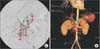
A 37-year-old woman was referred for multiple VAA found incidentally. Imaging studies revealed a total of five aneurysms. The origin of the celiac trunk was found to be stenotic with vascular collapse (Fig. 1A, B). An endovascular procedure was considered but deemed unsuccessful due to the artery location and tortuosity.
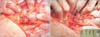
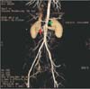
An exploratory laparotomy was performed. A saccular aneurysm with a broad base (a1) at the branching of the inferior pancreaticoduodenal artery was identified and circumferentially dissected (Fig. 2A). A round- and a straight-tipped Yasargil clips were used to exclude the aneurysmal sac and the circulation was preserved as identified by a palpable pulse (Fig. 2B). The saccular aneurysm (a2) in the inferior pancreatic artery was connected to a fusiform one (a3) that involved the entire arterial circumference. These two aneurysms were resected en bloc and the arterial stumps were anastomosed. The remaining two fusiform aneurysms, one in the inferior pancreatic artery (a4) and the other in the splenic artery (a5), appeared to involve only part of the arterial wall and were managed by the use of three more Yasargil clips. The operation took 11 hours, with the estimated blood loss of 400 mL. On pathologic examination, the aneurysm showed fibrosis, diffuse hyalinization and myxoid degeneration of the arterial wall, as evidenced by a marked loss of elastic fiber and smooth muscle (Fig. 3A, B). The postoperative recovery was uneventful, and she was discharged on the 17th postoperative day. An angiomesenteric CT performed at 36-month follow-up showed no evidence of recurrence or newly developed lesions, and flow restoration of the celiac axis. The inferior pancreaticoduodenal artery and the splenic artery remained intact, while the inferior pancreatic artery was not visualized (Fig. 4).
DISCUSSION
The etiology of VAA is unclear. Its known risk factors include arteriosclerosis, cystic medial necrosis, vasculitis, and autoimmune disorders [2]. Specifically, inferior pancreaticoduodenal artery and splenic artery aneurysms are commonly associated with pancreatic or biliary diseases and trauma [23]. The current patient did not have any of these factors except for a history of pregnancy [3]. The histopathological features of the resected artery of this patient were consistent with fibromuscular dysplasia [35], and the celiac artery stenosis could be explained by the same pathological process [6], although the possibility of congenital origin cannot be ruled out because she had not undergone a previous study [2]. Reports have suggested celiac trunk stenosis as the cause of VAA [678]. Recurrence or bleeding of the pancreaticoduodenal artery aneurysms tend to occur as a consequence of the associated celiac axis occlusion and the compensatory high blood flow in the pancreaticoduodenal artery [68]. On the other hand, there are studies that have reported successful treatment of aneurysms without correcting the celiac axis occlusion [7]. Percutaneous angioplasty has been widely used to correct celiac arterial stenosis [8]. In the current case, the guide wire could not pass through the stenosis of the celiac ostium. During 36-month follow-up, no aneurysm recurrence or newly developed lesions were observed, and interestingly, the celiac axis flow has been restored spontaneously. Chiou et al. [2] reported a case of inferior pancreaticoduodenal artery aneurysm associated with celiac artery occlusion, and they observed that after resection and reconstruction of the inferior pancreaticoduodenal artery, the Doppler signal of the gastroduodenal and splenic arteries were intensified. While the causal relationship between celiac artery stenosis and aneurysm development remains unclear, restoration of the celiac axis after aneurysm correction in our case suggested that the former may be the result and not the cause of the latter, contradictory to previous suggestions. For the celiac reconstruction to be due to back flow from the superior mesenteric artery (SMA), the celiac ostium should be occluded and not only stenosed, which was not the case. We speculate that the aneurysmal and cirsoid dilatation of the pancreaticoduodenal and inferior pancreatic arteries stole the celiac circulation and caused it to collapse and that after aneurysm clipping and resection, the increased resistance of the collateral arteries contributed to reconstitution of the celiac artery. More case-based evidences are required for a conclusion to be drawn.
There is no definite relationship between aneurysm size and risk of rupture, and there is no evidence that smaller aneurysms are safe [48]. One report stated that 17.6% of ruptured aneurysms were ≤1 cm [7]. Therefore, although unruptured or asymptomatic, these aneurysms should be treated because after rupture, mortality rates are 21%-100% [9]. Regarding VAA management, endovascular procedures provide an attractive alternative to open surgery with minimal morbidity and mortality [9]. We tried to catheterize the inferior pancreaticoduodenal artery aneurysmal sac via SMA. However, the guide wire was not advanced into the sac and the other aneurysm was fusiform in the middle portion of the inferior pancreatic artery, which was tortuous. Surgical options for VAA include ligation, aneurysmectomy, resection and anastomosis, graft interposition, and splenectomy in cases in which the aneurysm is located in the distal portion of the splenic artery [9]. We have found that intracranial aneurysm clips are easy to handle, relocatable, and can greatly reduce the operative time. Furthermore, since they are designed for intracranial use, these clips are biologically inert, nonferromagnetic, and have long durability [10]. Further studies using this technique intraabdominally are warranted.

 XML Download