

 PDF
PDF ePub
ePub Citation
 Print
Print


INTRODUCTION
Primary hyperparathyroidism (pHPT) is a common clinical endocrine disorder in western countries [1]. More recently, the prevalence of pHPT has increased compared to the past owing to development of biochemical tests [2]. Untreated pHPT can cause a cardiovascular disease, which becomes the cause of reduced survival rates [3].
pHPT can only be treated by the parathyroidectomy [4]. The guidelines from the Third International Workshop on the management of asymptomatic pHPT clarified the surgical indication of pHPT patients [5]. pHPT has been treated by traditional bilateral neck exploration (BNE) using intraoperative parathyroid hormone (IOPTH) with a cure rate of more than 95% [6]. The improvement of IOPTH assays and localization studies have enabled a minimally invasive parathyroidectomy (MIP) [7]. Many studies reported that the use of IOPTH enables the operators to find and excise additional abnormal parathyroids missed by localization study [89]. However, some authors have announced that IOPTH are useless in MIP if pathologic parathyroids are successfully localized [41011].
The aim of this study is to analyze the demographics, clinical presentations and surgical outcomes of the pHPT patients who received surgical management with versus without IOPTH and to investigate the difference of IOPTH drop rate according to time.
METHODS
Analysis of a database was performed on all patients who underwent parathyroidectomy for pHPT by a single surgeon from 2004 to 2013 at Korea University Guro Hospital. Familial disease and secondary hyperparathyroidism were excluded. Fifty-three patients with sporadic pHPT were eligible for the study.
Serum calcium, serum PTH, 24-hour urine calcium, and bone density were measured at the initial visit. All patients were evaluated with both sestamibi scan and ultrasonography preoperatively for localization. All ultrasonographies and sestamibi scans were reviewed by the surgeon and the radiologist. If pathologic parathyoids were found in sestamibi scan and the results of sonography was concordant with the results of sestamibi scan, patients were offered MIP without IOPTH (group 1, n = 39). If any localization studies failed to find the parathyroid or absence of concordance between the results of sestamibi scan and ultrasonography was found, patients underwent the MIP or the BNE using IOPTH (group 2, n = 14).
In group 1, all patients had received MIP, which was performed with a lower neck skin incision, and localized parathyroid was removed. In group 2, patients with enlarged parathyroid in sonography underwent MIP and we checked IOPTH to determine the need for more exploration. But patients in whom localized parathyroid failed to be found in both studies had BNE using IOPTH through a cervical collar incision. The baseline of IOPTH level was measured before parathyroid resection. Also, we measured the IOPTH at 5 and 10 minutes after excision (postoperative 5 minutes, postoperative 10 minutes). IOPTH assays were performed in all groups, but we could not check the results of IOPTH for patients of group 1 because the results of IOPTH were reported postoperatively. The IOPTH level was determined with an Elecsys 2010 apparatus (Roche Diagnostics Co., Indianapolis, IN, USA).
In group 2, the indication of successful operation was the decline of IOPTH over 50% at any time. Routine frozen biopsy of parathyroids was not carried out. If the intrathyroid nodule was found in preoperative ultrasonography, partial thyroidectomy was combined.
Curative parathyroidectomy was defined as maintaining normocalcemia for at least 6 months after surgery. The criteria of normocalcemia was from 8.5 to 10.2 mg/dL. Persistence of hyperparathyroidism was defined as persistent hypercalcemia within 6 months of the initial operation. Recurrence of hyperparathyroidism was defined as elevated calcium at or beyond 6 months postoperatively. Data were obtained by reviewing hospital records. All patients were followed for at least 6 months. Follow-up information was obtained through patient visits or phone contact.
Stastistical evaluation was performed using IBM SPSS Statistics ver. 20.0 (IBM Co., Armonk, NY, USA). We analyzed the difference of IOPTH drop rate between cured group and noncured group. A P-value less than 0.05 was considered as statistical significance in all analyses.
RESULTS
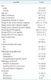
Among 53 patients, 34 (64.2%) were female and 19 (35.8%) were male. Mean age was 52.8 ± 15.5 years. At the time of the first presentation, 35 of the 53 patients (66.0%) were asymptomatic. 10 patients (18.9%) had symptoms related to ureteral stones. Neurological symptoms such as stupor and depressive mood change were revealed in three patients (5.7%). Four patients (7.5%) complained of bone related symptoms; All of them had a history of fracture. One patient who came to the emergency room was diagnosed with acute pancreatitis with hypercalcemia (Table 1).
Table 2 shows the demographic, laboratory, clinical and surgical data of all patients. The mean preoperative serum calcium was 11.6 ± 1.1 mg/dL, the serum ionized calcium was 5.9 ± 0.6 mg/dL, and the 24-hour urinary calcium excretion was 254.3 ± 174.6 mg/day. Patients diagnosed with osteoporosis (T score < -2.5) were 24 (45.2%) in the preoperative bone density test. The mean T score of patients was -2.5 ± 1.0.
The sestamibi scan results of only 39 patients (73.6%) corresponded with their surgical findings. But, preoperative ultrasonography findings of 48 patients (90.6%) showed correlation with operative findings. All 5 patients who failed localization initially received BNE.
Combined thyroidectomy was performed for 26 patients (49.1%). Of them, 5 patients (19.2%) with no preoperative thyroid nodule had a partial thyroidectomy owing to suspicious operative findings, and no pathological diagnosis was confirmed in all. The others had a thyroidectomy due to combined thyroid nodules. Fourteen of 26 patients (56.0%) had nodular hyperplasia, 6 patients (24.0%) had papillary thyroid carcinoma, and 5 patients (20.0%) had no pathological diagnosis finally.
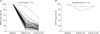
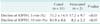
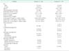
The mean preoperative basal PTH was 228.4 ± 251.6 pg/mL. The decline of PTH at 5 minutes and 10 minutes postoperative was 75.2% ± 14.9% and 84.9% ± 8.6%, period of over 6 months in cured patients. On the other hand, that of noncured patients at 5 minutes and 10 minutes was 17.2% ± 9.7% and 8.2% ± 2.2%, respectively. There was a significant difference for the decline of IOPTH between cured and noncured patients (Table 3). There were two patients who were not satisfied with the 50% drop of IOPTH at 5 minutes after excision. But their IOPTH at 10 minutes decreased below 50% compared to basal IOPTH and the patients were cured successfully from their pHPT (Fig. 1).
The overall cure rate of 39 patients in group 1 versus 14 patients in group 2 was 94.9% versus 100%, but there was no stastistical difference. Also, there was no significant differences between the two groups in sex, age, preoperative T score, 24-hour urine calcium, serum calcium, basal PTH level, postoperative PTH level, combined thyroidectomy or pathology of thyroid and parathyroid (Table 4).
During the follow-up respectively, 51 patients (96.2%) achieved normocalcemia. There was no recurrence, but persistence of pHPT was shown in 2 patients of group 1. Pathology of excised parathyroids was consistent with adenoma in 41 of 53 patients (77.4%) and hyperplasia in 10 of 53 patients (18.9%). There is no complication such as hypocalcemia, hoarseness in all patients.
DISCUSSION
Since the first parathyroidectomy eighty years ago, the treatment of pHPT had been BNE, which showed excellent results by experienced surgeons [1213]. Recently, this surgical standard has changed focus to parathyroidectomy due to improvements in preoperative imaging and intraoperative modalities such as IOPTH [14]. Minimally invasive surgeries for pHPT are associated with short operative time, rapid postoperative recovery and low complication rates [1516]. Especially, many studies have reported that IOPTH monitoring helps to determine the need for complete resection of hyperfunctioning parathyroid in both MIP and BNE [1718]. In this study, we compared the surgical outcomes in MIP between groups 1 and 2 (without IOPTH versus using IOPTH). All of two persistent cases occured in only group 1 using no IOPTH. There was a significant difference between the two groups regarding surgical failure (P < 0.01).
Measurement of IOPTH in parathyroidectomy was first described by Nussbaum et al. [19]. Compared with baseline PTH level, a decline of 50% or more in IOPTH means that hyperfunctioning parathyroid tissue has been excised [20]. There is an argument about the time of PTH measurement after excision [21]. Barczynski et al. [22] had reported that when they analyzed the Halle, Miami, Rome, and Vienna IOPTH Criteria in MIP, each criteria had a distinguishing limitation. Above all, the Miami Criteria, i.e., a decline in IOPTH of 50% or more at 10 minutes after excision, showned acceptable sensitivity and specificity in detecting multiple gland disease [21]. We checked the IOPTH at 5 and 10 minutes, and two patients had a decline of IOPTH of 50% or less at 5 minutes. But they finally showed a decline of IOPTH of 50% or more at 10 minutes. Using the Miami Criteria, we could predict the cure of pHPT in all patients.
Symptomatic pHPT is rare today, but renal and ureteral stone still occurs in 6% to 15% of cases [23]. In our study, 10 patients (18.9%) suffered from nephrolithiasis, which are slightly higher incidence rates than that of other studies. Moreover, symptomatic pHPT in our study was 18 patients (34.0%), and it is more common than that in the West. The mean age was 52.8 ± 15.5 years and the ratio of sex was 19 : 34 (male:female). It was not different from previous studies [24].
Identification of pathologic parathyroid is important for deciding on how to operate. Ultrasonography successfully identifies a solitary adenoma in 61%-92% of patients, and when combined with sestamibi has been found to have greater than 90% sensitivity [25]. Sestamibi scan is a useful modality for finding abnormal glands, and it has been found to be 50%-91% sensitive in localization of pathologic glands. But many studies reported variation of sensitivity [2627]. Several studies had reported that sestamibi scan could not detect a smaller parathyroid due to low resolution [28]. We found that sensitivity of sestamibi scan was only 73.6%, but that of ultrasonography was 90.6%.
Coexistence of thyroid nodules is found in 20%-60% of patients with primary HPT [29]. Those findings should be evaluated preoperatively because there are possibilities for unsuspected thyroid cancer or intrathyroid parathyoid [30]. In our series, combined thyroidectomy was performed in 26 patients (49.1%). Among them, six patients had a thyroid carcinoma and 15 patients were diagnosed with nodular hyperplasia. But there was no intrathyroid parathyroid.
There are some limitations to our study. First, due to a small number of patients, we could not analyze the variables that influenced persistent or recurrent disease. With regard to this, additional multicenter trials should be carried out. Second, our study has the limitation of a single institution study. It may be subjective to referral bias.
In conclusion, even though the localization studies were successful, IOPTH monitoring is essential to avoid surgical failure in MIP. Especially, the Miami Criteria is a proper tool in evaluation of successful parathyroidectomy.
 XML Download