

 PDF
PDF ePub
ePub Citation
 Print
Print


INTRODUCTION
An acute interruption in the forward flow of the luminal contents in the gastrointestinal system accounts for acute mechanical intestinal obstruction (AMIO) [1]. This entity was first diagnosed and treated by Hippocrates. The first documented treatment was performed by Praxagoras in 350 BC, when he created a decompressive fistula between the skin and the bowel of a patient with AMIO [2].
Although the most common reason for AMIO was incarcerated hernia in the first decades of the 20th century, postoperative adhesions have currently taken first place as an etiological factor due to increased number and variety of therapeutic intra-abdominal surgical interventions [3].
Any surgical procedure in the peritoneal cavity results with intra-abdominal adhesions, which are strands or membranes of fibrous tissue that can be attached to various organs, sticking together and leading to AMIO [4]. Abdominal adhesions, which can begin forming within a few hours after an operation, represent the most common cause of intestinal obstruction being responsible for 60%-70% of AMIO cases [5].
AMIO remains a leading cause of hospital admission [4,6]. The morbidity and financial cost of AMIO due to adhesions are compounded by the recurrent nature of the disease [7]. Mortality rates may vary from 3% in simple obstructions, to 30% in cases harbouring strangulation, or presence of comorbidities [2]. Recurrence has been reported to take place in 12% of patients after conservative treatment, and in 8%-32% of patients after surgical treatment [2].
Although it has been reported in high-volume retrospective and prospective studies that 60%-80% of the patients are treated conservatively while 20%-40% are treated surgically, controversy still exists concerning the type of treatment and timing of operation if surgery is required [1,4,6]. For this reason, research continues in the quest of a surgeon to rule out the predictive factors indicating intestinal ischemia [4].
Our aim was to investigate the predictive factors indicating intestinal ischemia in patients with the diagnosis of AMIO due to adhesions, in order to display the surgical indications for its operative treatment.
METHODS
This study retrospectively evaluated the data of all patients who were diagnosed with and treated for AMIO due to adhesions at a single-center institution (Istanbul Medeniyet University School of Medicine, Department of General Surgery) between January 2009 and January 2014.
Approval for this study was obtained from the Ethics Committee of the Istanbul Medeniyet University Goztepe Training & Research Hospital. Signed informed consent forms were obtained from all patients who were included in the study.
All patients who had been admitted to the hospital with documented AMIO, and having had a past history of one or more intra-abdominal operations or a history of a disease causing intra-abdominal inflammation were considered to possess intra-abdominal adhesions. Following the exclusion of the patients with AMIO due to other reasons (such as tumoral obstruction, incarcerated hernia, etc.), all patients under the diagnosis of "AMIO due to adhesions" were included in the study. The patients were separated into two groups according to their modality of treatment; patients who underwent surgery (operative treatment group, group S), and patients who were treated conservatively (nonoperative treatment group, group C).
Clinical and epidemiological data on file, information on admission, and the diagnostic work-up (age, sex, findings on physical examination, laboratory and radiological assessment) were analyzed and correlated with the treatment modalities and surgical outcomes (i.e., the patients' early postoperative follow-up parameters, such as the time to the postoperative first flatus, the first defecation, and the initiation of oral intake, the duration of the hospital stay, complications, morbidity, and mortality) of the two groups.
WBC (normal range, 4,000-10,000/mm3), CRP (normal range, 0-0.5 mg/dL), Urea (normal range, 17-43 mg/dL), Creatinine (normal range, 0.7-1.2 mg/dL), Sodium (Na; normal range, 134-146 mEq/L), and Potassium (K; normal range, 3.5-5.2 mEq/L) levels were recorded and analyzed as the laboratory findings of major importance.
In cases having had a plain abdominal radiography (PAR), patients were separated into three groups as "mild intestinal obstruction", "severe intestinal obstruction", and "colonic obstruction" according to the number and location of the air-fluid levels, and the diameter of the dilated loops. The term "severe intestinal obstruction" was used in cases with ≥36 mm of a maximum diameter of the dilated intestinal segments, if this intestinal image exceeded the 50% diameter of the largest colonic segment that could be visualized, and if the number of dilated intestinal segments was more than 2.5 times the number of the normal segments [8].
In patients having had an abdominal CT scan, the findings of AMIO and its level, and the signs of either intestinal, or colonic ischemia, or strangulation (intestinal inflammation, thickened bowel wall, ascites, a trilaminar appearance of the bowel wall known as the target sign, poor or absent enhancement of bowel wall on intravenous contrast enhanced scans, pneumatosis intestinalis, gas in mesenteric or portal veins, twisting of the mesenteric vasculature defined as the whirl sign, tortuous engorged mesenteric vessels, mesenteric hemorrhage, increased attenuation of bowel wall on noncontrast scans) were documented [9].
The patients in the surgically treated group (group S) were also separated into two groups according to their intraoperative findings; as the group with the presence of strangulation (bowel ischemia group, group I), and the group with operative findings of adhesions without any evidence of bowel ischemia (adhesion only group, group A).
The patients in group S were also separated into two groups according to the time period between admission and operation, as the group of patients operated on within the first three days of admission (early operation group, group E), and the group of patients operated on after the end of the first three days of admission (late operation group, group L).
The NCSS (Number Cruncher Statistical System) 2007 & PASS (Power Analysis and Sample Size) 2008 Statistical Software (Kaysville, UT, USA) program was used for statistical analysis. Additional to the descriptive statistical methods (mean, standard deviation, median, frequency, ratio), the Student t-test was used to compare numerical quantities of parameters with normal distribution. The Mann-Whitney U test was used for the comparison of two groups with any maldistribution of parameters. Fisher exact test and Yates' continuity correction were used for the qualitative comparisons of the data. Differences were considered statistically significant when P < 0.05.
RESULTS
The medical files of 580 patients with the diagnosis of AMIO who had been treated either surgically or conservatively in the Department of Surgery of our institution between January 2009 and January 2014 were investigated, and, according to the exclusion criteria, 252 patients having had the etiology of adhesions were included in the study.
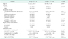
The total study group consisted of 113 women (44.8%), and 139 men (55.2%). The mean age was 62.79 ± 18.08 years (range, 20-98 years). The surgical (operative) treatment group (group S) consisted of 50 patients (19.8%) while the remaining 202 patients (80.2%) were in the conservative (nonoperative) treatment group (group C). The demographic data of group S, and group C was similar (Table 1).
The prehospital symptomatic period (the time from the onset of symptoms till the time of admission) was significantly longer in group S (P < 0.01). Among all signs, presence of fever was found to be statistically significant in group S (P < 0.01). The comparison of the symptoms & signs at first admission between group S, and group C is summarized in Table 1.
The laboratory tests revealed significantly elevated CRP levels in group S (P < 0.01). The comparison of the laboratory tests between group S, and group C is shown in Table 1, as well.
Among all 252 cases, 248 patients (98.4%) had a past history of at least one or more intra-abdominal surgical interventions. 164 patients (65.1%) revealed a history of a single former operation. Additionally, 62 patients (24.6%) had undergone two, 16 (6%) had undergone three, and 6 (2.4%) had undergone four former surgical interventions.
When these former interventions were evaluated, it was found that a total of 341 prior operations were performed in our study group. These interventions included 51 gynecological procedures (20.2%), 49 appendectomies (19.4%), 48 colorectal resections (19.0%), 43 gastric surgical procedures (17.1%), 43 hepatobiliary operations (17.1%), 37 operations (14.7%) for prior adhesive AMIO, 34 abdominal wall hernia operations (13.5%), 16 diagnostic laparotomies (6.3%), 14 urological operations (5.6%), and 6 intestinal surgical procedures (2.4%).
The four patients (1.6%) not having undergone any operations consisted of three men and one woman. She revealed a past history of severe pelvic inflammatory disease. Her intraoperative findings were dense adhesions between the small bowel segments, and the adnexal structures. One of the three men had a past medical history of tuberculous peritonitis, and his intraoperative findings were diffuse adhesions of the bowel accompanied by peritoneal nodule formations, the biopsies of which ended up to be defined as caseous necrosis after the pathological evaluations. The other two men revealed a history of inflammatory bowel disease, confirmed intraoperatively as well.
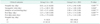
PAR was order for all patients at first admission. According to the PAR images the frequency of "mild intestinal obstruction" was significantly lower in group S (P < 0.01). On the other hand, the incidence of PAR findings indicating "severe intestinal obstruction" was significantly higher in group S than group C (P < 0.01). Additionally, an abdominal CT scan was performed in 146 patients (57.9%). The incidence of CT findings indicating bowel obstruction is significantly higher in group S (P < 0.01). Indirect images of strangulation was detected in 10 patients (20%) in group S, but none in group C. The comparisons of the radiological evaluations between the two groups are summarized in Table 2.
In group S, two patients (4%) were operated on laparoscopically while the remaining 48 (96%) were operated on via the conventional (open) technique. Adhesiolysis was performed in all cases in group S. The cases that had intraoperative findings of strangulation, or perforation, had undergone either segmental small bowel or segmental colon resections with or without the creation of a stoma. Twelve patients (24%) underwent a segmental small bowel resection, and 4 patients (8%) underwent a segmental colon resection. An ileostomy was needed to be created in 3 patients (6%), while the creation of a colostomy was required in 3 patients (6%).
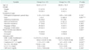
The duration of hospital stay in group S was found to be significantly longer than in group C (P < 0.01). Hospital stay was >3 days in 82% of the patients in group S. On the other hand, the time period for the initiation of oral feeding, the first flatus and the first defecation was significantly longer in group S (P < 0.01). The outcomes of the two different modalities of treatment are shown in Table 3.
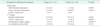
The patients in the surgically treated group (group S) were separated into two groups according to their intraoperative findings; the cases in which bowel ischemia was observed (group I), and the group with operative findings of adhesions without any bowel ischemia (group A). Group I consisted of 19 patients (38%), where as 31 patients (62%) were in group A.
When the demographic data was compared between groups A and I, it was detected that elderly patients were more prone to strangulation (P < 0.05) (Table 4). However, this finding was not affected by the gender of the patients.
The comparison of the symptoms & signs between groups I and A revealed that the incidences of fever, and rebound tenderness were significantly higher in cases with the presence of strangulation (Table 4).
When laboratory tests were compared between groups I and A, it was detected that the urea & creatinine levels were significantly higher in group I (P < 0.05) (Table 4).
Findings of "severe intestinal obstruction" at PAR were found to be significantly related with strangulation (P < 0.05). Indirect signs of bowel ischemia detected via preoperative abdominal CT scan revealed a statistically significant relation with intraoperative findings of strangulation (P < 0.01). Other CT findings such as the level of obstruction, or the presence of intra-abdominal fluid did not show statistical importance (P > 0.05). The comparisons of the preoperative radiological evaluations between groups I and A are summarized in Table 5.
In group S, the mean duration between admission to the hospital and the surgical intervention was calculated to be 2.80 ± 3.34 days (range, 1-16 days).
The patients in group S were also separated into two groups according to the time period between admission and operation, as the group of patients operated on within the first three days of admission (group E), and the group of patients operated on after the end of the first three days of admission (group L).
Group E consisted of 37 patients (74%), while 13 patients (26%) were in group L. No significant differences were detected between groups E and L in concern of preoperative tachycardia (P = 0.301), intraoperative findings of strangulation (P = 0.199), complications (P > 0.999), or mortality rates (P = 0.662) (Fisher exact test; P > 0.05).
DISCUSSION
AMIO, is a common reason for emergency surgery worldwide [4,6]. The morbidity, and mortality associated with AMIO continue to be significant. Mortality rates may vary from 3% for simple obstructions, to as great as 30% for cases with the presence of strangulation accompanied by comorbidities [2]. As a major etiological factor, adhesions resulting from prior abdominal surgery account for 40%-80% of AMIO cases, and this wide variation in the incidence of adhesive obstruction alters with different referral patterns, community settings, racial cultures, and countries [2]. Recurrence rates have been reported to be 12% after a successful primary conservative treatment, and 8%-32% following surgery for AMIO due to adhesions [2].
Lower abdominal surgeries including appendectomies, colorectal surgery, gynecologic procedures, and hernia repairs confer a greater risk of adhesive AMIO [1]. In two different series, it was stated that women represented approximately 60% of all cases [6,10]. According to the results of another prospective study on 124 patients conducted in our country, the female ratio was found to be 45% [11]. In our total study group of 252 patients, 55% consisted of men, and 45% were women. No statistical significance was detected in concern of gender between either groups S andC, or groups A and I (P > 0.05, and P > 0.05, consecutively).
Although it may take place in any decade of life, the incidence of AMIO increases with age. In different studies, the mean age was found to range between 55 and 65 [11,12,13]. The mean age was 62.79 ± 18.08 years (range, 20-98 years) in our study group. When this parameter was compared between groups A and I, it was detected that the elderly patients were more prone to strangulation (P < 0.05) (Table 4).
A thorough medical history to be obtained, and a complete physical examination is very important in AMIO cases. When supported by a PAR, these simple steps are usually adequate for the primary diagnosis. Abdominal pain, nausea & vomiting, stoppage of the passage of flatus or stool, abdominal distension constitute the classical symptoms, and these symptoms may vary according to the time period, level, and severity of AMIO [4,6]. In their study of 300 patients, Cheadle et al. [13] stated that abdominal pain (92%), vomiting (82%), abdominal tenderness (64%), and distension (59%) were the most frequent symptoms & signs. In another series of AMIO, Perea Garcia et al. [14] reported that the most prevalent symptoms & signs were vomiting (77%), colicky pain (68%), stoppage of flatus or defecation (52%), and abdominal pain (12%), respectively. The symptoms & signs of the present study group are listed in Table 1.
Bowel obstruction may be presented in either an acute form, or chronic relapsing symptoms. The urgency of diagnosis and treatment differ between these two groups. The time of admission to hospital may alter according to the severity of the symptoms [2]. There may be a delay in the definition of symptoms, and admission to the hospital in elderly patients with different levels of cognitive decline, as well [4]. Markogiannakis et al. [6] calculated the prehospital symptomatic period to be 2 days in their series. In our study, the prehospital symptomatic period was 4 days. This longer delay period may be due to the particular cultural differences in our country.
Adhesive AMIO may be related with congenital or acquired reasons. Acquired etiologies may take place because of a prior operation, or a former inflammatory process. Sixty-five to ninety percent of the patients diagnosed with adhesive AMIO have a history of one or more prior operations [11]. In a prospective observational study on 150 patients published in 2007, it was reported the vast majority of the cases had undergone one operation (n = 70, 72.1%), 18 (18.6%) had two, and 9 (9.3%) had three operations [6]. Among all 252 patients in the present study group, 4 subjects (1.6%) didn't have an operative history, and the majority of the remaining subjects (65.1%) revealed a history of a single former operation. Additionally, 24.6% of the subjects had undergone two, and 6% had undergone three former surgical interventions.
Adhesions are more frequent following operations performed in the lower abdominal cavity and pelvis, such as obstetric & gynecologic procedures, colorectal resections, and appendectomies because of the free movement of the small bowel in the pelvis and the formation of stronger adhesions in this location compared to the upper abdominal cavity [15]. In their retrospective study of 123 patients carried out in 2004, Kossi et al. [15] reported that the past history of previous operations of their patients revealed rates of 32.4% colorectal, 27.8% upper abdominal, 19.9% gynecological, 8.5% middle abdominal, 5.1% abdominl wall, 4.5% urological, and 1.7% undefined interventions. In the related studies, adhesive AMIO was considered more likely to take place after operations of the lower abdominal cavity and pelvis, especially in which the peritoneal surfaces are widely exposed or damaged. It was reported that adhesive AMIO rates were 1%-10% after appendectomy, 7% after conventional cholecystectomy, 10%-25% after bowel surgery, and 17%-25% after proctocolectomy [16]. In our series, gynecological procedures (20.2%), appendectomies (19.4%), and colorectal resections (19.0%), followed by gastric (17.1%) and hepatobiliary procedures (17.1%) were found to be the most prevalent prior surgical interventions in cases of adhesive AMIO.
Controversy still exists for the choice and timing of treatment in AMIO due to adhesions. Surgical treatment may result with recurrence because of new adhesion formations, and nonoperative treatment does not resolve the primary reason of obstruction. According to the Bologna Guidelines, a nonoperative approach may be the treatment of choice in cases without the presence of clinical, laboratory, or radiologic findings of strangulation [4]. Patients treated nonoperatively have shorter hospital stay, but higher recurrence rates, and shorter time to readmission [4,10]. Recurrence rates are lower in surgically treated adhesive AMIO patients compared to conservative treatment, but the surgical indications will remain unchanged for a newly developed episode of adhesive AMIO [7,17,18]. The need for operative intervention was detected to be 14%-44% in other reports, and the duration of hospital stay with nonoperatively treated patients was 5 to 7 days, where as this time period was 11 to 19 days with surgically treated patients [15,19,20]. In a series of 27,046 patients published in 2012, it was detected that 18% of the cases required surgery with a mean length of hospital stay of 8.51 days, and the remaining 82% were treated conservatively staying a mean of 4 days [10]. In the present study, 50 cases (19.8%) were treated surgically (group S) while the remaining 202 (80.2%) patients received nonoperative treatment (group C) with a mean hospital stay of 8.92 days for group S, and 4.74 days for group C revealing a significant difference between the two treatment groups (P < 0.01) (Table 3).
On one hand, the presence of elevated WBC counts is not accepted to be an individual predictive factor for strangulation, and on the other hand, WBC counts may sometimes be normal in the presence of strangulation [21]. When combined with WBC counts and CT findings, the detection of CRP levels was found to be significantly indicative for surgical treatment [22]. In our study, fever was significantly more prevalent (P < 0.01), and CRP levels were significantly higher (P < 0.01) in group S. Other laboratory findings including WBC, Urea, Creatinine, Na, and K did not reveal any statistical significance between groups S and C (P > 0.05) (Table 1).
In cases of AMIO, it is not always easy to define the presence of strangulation (bowel ischemia). Some studies reveal that despite close and careful clinical evaluation, in conjunction with laboratory and radiologic studies, a preoperative diagnosis of bowel strangulation can not be made or excluded reliably by any known parameter, combinations of parameters, or by experienced clinical judgement [3,6,10,23]. Persistent abdominal pain, fever, tachycardia, abdominal tenderness, rebound tenderness, muscular resistance are known as predictive factors for strangulation in AMIO, and the risk of strangulation was determined to be 82%, and 100%, with the presence of three, and four of these signs, consecutively [24]. The statistical comparisons of groups I and A in our series also revealed that fever and rebound tenderness were significant predictive signs for strangulation (P < 0.01 and P < 0.05, consecutively) (Table 4).
In cases of bowel obstruction, different levels of fluid - electrolyte imbalances may take place. High serum urea, and creatinine levels are important parameters that display dehydration which may be a result of sepsis or multiorgan dysfunction syndrome due to strangulation, or perforation. Additionally, alkalosis with hypochloremia and hypopotassemia may accompany the clinical state. Fluid resuscitation must be carried out for these patients with close monitoring of the urinary output [4,25]. In this study, the urea and creatinine levels were significantly elevated in group I compared to group A displaying the frequency of dehydration in the presence of strangulation (P < 0.05 and P < 0.05, consecutively). Other laboratory findings including WBC, CRP, Na, and K did not reveal any statistical significance between groups A and I (P > 0.05) (Table 4). Thus, CRP analysis was considered to be useful for the decision of surgery, but not significantly predictive for strangulation.
Since PAR is a simple and inexpensive imaging tool, it remains the initial choice for radiologic evaluation in cases of AMIO; however, in conjunction with the clinical examination this modality is diagnostic in only 50%-60% of cases [8,26]. Concerning the PAR findings of our series, mild intestinal obstruction rate was significantly higher in group C, while severe intestinal obstruction rate was detected to be significantly higher in group S (P < 0.01 and P < 0.01, consecutively) (Table 2). On the other hand, the prevalence of severe intestinal obstruction detected via PAR was found to be significantly higher in group I when compared with group A (P < 0.05) (Table 5). According to these results, PAR findings of severe intestinal obstruction was considered to be significantly indicative for surgical treatment, and was related with a possible presence of strangulation.
CT has a sensitivity of 81%-94% and a specificity of 96% for diagnosing AMIO [12,27]. Beyond any doubt, the most important information that CT can provide the surgeon is whether there is an associated strangulation [9]. The sensitivity of contrast-enhanced CT for intestinal ischemia has been reported to be as high as 90% [26]. CT was used for the advanced radiological evaluations of 146 patients (57.9%) in the present study group. The incidence of CT findings indicating bowel obstruction is significantly higher in group S (P < 0.01) (Table 2). Indirect signs of bowel ischemia detected via CT scan revealed a statistically significant relation with intraoperative findings of strangulation (P < 0.01) (Table 5). According to these results, CT findings of bowel obstruction was considered to be significantly indicative for surgical treatment, and the indirect signs of bowel ischemia detected via CT were significantly specific.
Usually nonoperative treatment, in the absence of signs of strangulation or peritonitis, can be prolonged up to 72 hours of adhesive AMIO [4]. If there is no resolution within 3-5 days, surgery should be considered [28,29]. If ileus persists more than 3 days and the nasogastric tube drainage volume on day 3 is >500 mL, surgery for AMIO is recommended [4]. With close monitoring and in the absence of signs suggestive of complications, an observation period even longer than 10 days before proceeding to surgical intervention appears to be safe [30]. However, at any time, if onset of fever and leukocytosis greater than 15,000/mm3 (predictors of intestinal complications) are observed, then conservative treatment should be discontinued and surgery is recommended [4]. In group S, the mean duration between admission to hospital and surgical intervention was calculated to be 2.80 ± 3.34 days (range, 1-16 days). The patients in group S were also separated into two groups according to the time period between admission and operation, as the group of patients operated on within the first three days of admission (group E), and the group of patients operated on after the end of the first three days of admission (group L). Group E consisted of 37 patients (74%), while 13 patients (26%) were in group L. Although the number of patients with intraoperative findings of bowel ischemia was higher in group E when compared to group L, this issue did not reveal significance as results of the statistical analysis (P > 0.05). No significant differences were detected between groups E and L in concern of preoperative tachycardia, postoperative complications, or mortality rates, either (P > 0.05).
As a conclusion, it can be stated that longer prehospital symptomatic period is related with a tendency for surgical treatment, and elderly patients are more prone to strangulation when adhesional AMIO takes place. The statistically significant parameters of our series, which are fever, rebound tenderness, elevated urea & creatinine levels, severe intestinal obstruction findings in PAR, and indirect findings of strangulation in CT, are important indicators of a possible bowel ischemia in cases of AMIO due to adhesions. CRP detection was considered to be useful for the decision of surgery, but not significantly predictive for strangulation. Thus, these findings in conjunction with clinical experiences may guide the surgeon in the decision making of an emergency surgical intervention.

 XML Download