

 PDF
PDF ePub
ePub Citation
 Print
Print


INTRODUCTION
The right internal jugular vein (IJV) is the preferred site for hemodialysis catheter insertion. The right IJV has a straight course to the superior vena cava. Especially, the use of the IJV can reduce the potential complications of subclavian-based access, such as thrombosis/stenosis of the subclavian vein and pinch-off syndrome [1,2]. Hence, the right IJV is generally the first choice site for central catheterization. A recent widespread conception is that ultrasound-guided catheter insertion is a mandatory method [3]. The operator can determine the best choice of the vessel. A recently published meta-analysis demonstrated that ultrasound guidance clearly reduces the number of attempts and the incidence of procedure failure [4].
Some techniques have been introduced for ultrasound-guided central venous catheterization. Among them, short-axis lateral in-plane technique is considered to be the most useful technique for IJV access [5]. Therefore, we used the "short-axis, lateral in-plane technique" for the insertion of a large bore cuffed tunneled dual lumen catheter for hemodialysis. The aim of this paper is to describe the efficacy of "short-axis lateral in-plane technique" for hemodialysis catheterization with detailed consideration of technical points.
SURGICAL TECHNIQUE
A 59-year-old man was suffering from hypertension, diabetes, and cerebral infarction. The patient was diagnosed with end stage renal disease, and hemodialysis was initially started with prosthetic arteriovenous graft at the left forearm. After 2 years, the prosthetic arteriovenous graft was thrombosed and it failed as a salvage procedure. The patient was referred for a new vascular access creation. As a bridge to hemodialysis before new access creation and maturation, a cuffed tunneled two lumen hemodialysis catheter insertion via the right IJV was planned.
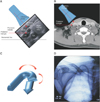
In the supine position under local anesthesia, the head was turned slightly to the left side and the sternocleidomastoid (SCM) muscle popped out of the neck. For ultrasound-guided puncture, the Doppler ultrasound probe was wrapped in a sterile cover. The right IJV and carotid artery were identified using Doppler ultrasound. The diameter of the IJV was 14 mm, being anterolateral in relation to the carotid artery. The entry site of the needle was lateral to the lateral margin of the SCM muscle in the posterior triangle and it was directed towards the IJV at an angle parallel to the horizontal plane (Fig. 1A, B). Cannulation of the IJV was achieved on the first attempt of puncture. The vein was entered within 5- to 7-cm depth of the needle. Retraction of the syringe plunger produced a flush of dark red blood when the IJV was entered. After puncture of the superior lateral wall of the IJV, a J-tip steel guidewire was passed through the needle into the vein. The skin over the guidewire exit site was minimally incised to accommodate at least the diameter of the catheter. An incision was made on the planned catheter exit site below the midclavicle. The catheter (HEMO-FLOW, Medcomp Components Inc., Harleysville, PA, USA) was advanced subcutaneously from the catheter exit site to the guidewire exit site. Care was taken to ensure that the tunnel provides a gentle curve to the catheter from the catheter exit site to the guidewire exit site. After dilating the vein, the dilator/sheath combination was placed over the wire. The dilator was removed, the catheter was advanced through the sheath, and the sheath was peeled away. The position of the tip of the catheter was checked. There were no immediate postprocedural complications. The time to catheter placement was 15 minutes.
DISCUSSION
The three essential components for acquiring images via ultrasound-guided needle approach are the vessel, ultrasound probe, and the puncture needle. We can obtain four different techniques according to the positional relationship of these three components. Ultrasound probe can be placed either in the "short axis" (Probe is perpendicular to the course of the vein) or in the "long axis" (Probe is parallel to the course of the vein) of the IJV. Additionally, the puncture needle can have two trajectory directions, "in-plane" (Needle is running parallel to the long axis of the probe) or "out-of-plane" (Needle is at right angles to the long axis of the probe) of the ultrasound probe [5].
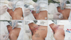
In the "short-axis out-of-plane" view, the operators have a very limited view of the needle tip and an adjustment of the probe is needed to have the needle tip aligned with the ultrasound beam. The operators have to coordinate the needle and ultrasound probe movements (Fig. 2A). In the "long-axis in-plane" view, the whole course of the IJV can be seen with the in-plane technique. However, surrounding structures (carotid artery, thyroid, lymph nodes) cannot be visualized. The needle insertion level is higher than that in the other technique as long as the probe length. Although the probe is positioned on the patient's neck with its caudal edge in contact with the clavicle, the needle would be inserted from the cranial edge of the ultrasound probe, away from the clavicle (Fig. 2B). In the "long-axis out-of-plane" view, the operators cannot see the whole course of the needle and also do not obtain complete information on the surrounding tissue. Thus, the risk of unwanted accidental puncture of surrounding structures may be high (Fig. 2C). In the "short-axis lateral in-plane" view, the operators can visualize the surrounding structures simultaneously with visualization of the whole length of the needle. This allows the operator to avoid iatrogenic puncture complications, such as arterial puncture and pneumothorax. Additionally, no adjustment of the probe is required during the procedure. Probe is positioned in transverse orientation just above the clavicle. The needle is inserted at the lateral edge of the ultrasound probe. Hence, the needle can be inserted nearly from the clavicle. This means that the inserted catheter has a more smooth curvature over the clavicle (Fig. 2D). The ultrasound-guided short-axis lateral in-plane technique for IJV cannulation is thought to be the most effective among the four methods. This method should be considered as the first-line technique [5].
For the exact skin exit site of the catheter, Rossi et al. [5] considered that a lateral, 30° access at the Sedillot's triangle is the safest and most effective percutaneous technique to reach the IJV. The SCM muscle splits into the sternal and clavicular heads; the division of the muscle with the clavicle creates a space called "Sedillot's triangle. For this method, the catheter should have two mixed curvatures; the catheter is first bent at the horizon and is directed downwards from the horizontal line (Fig. 1A-C). However, the IJV usually lies beneath the SCM muscle. Needle direction from the posterior triangle parallel to the horizontal line may need just one curvature (Fig. 1A, B, D; Fig. 2E, F). Hemodialysis catheter usually provides high blood flow in a back and forth fashion. Because a lesser number of angulations may lead to good flow rates and catheter function, we thought that skin puncture site in the neck at the posterior triangle is better than the Sedillot's triangle. Using this approach, we can reduce the possible complications of pinching and kinking of the catheter.
CONCLUSION
The IJV with surrounding structures can be visualized in the short axis view, while the puncture needle can be introduced using the "in-plane" technique allowing visualization of the whole length of needle. This technique is feasible for inserting a large bore cuffed tunneled dual lumen catheter for hemodialysis. The authors recommend the needle approach at the posterior angle of the neck to minimize the curvature of the catheter, especially for hemodialysis catheterization which needs high flow for effective hemodialysis.
 XML Download