

 PDF
PDF ePub
ePub Citation
 Print
Print


INTRODUCTION
Portal vein thrombosis (PVT) with cavernous transformation of the portal vein (CTPV) represents a surgically demanding challenge in living donor liver transplantation (LDLT) [1]. Although various surgical techniques and approaches to PVT has been introduced to overcome PVT, the absence of portal flow remains a surgically demanding challenge in the setting of LDLT because it has an obvious influence on surgical complexity, morbidity and mortality rates [1,2,3]. Furthermore, portal vein (PV) reconstruction is a very difficult procedure in LDLT with extensive PVT with CTPV in the setting of decreasing portal hypertension and providing adequate portal inflow [3,4]. Therefore, most transplant surgeons have hesitated to perform LDLT in cases with diffuse PVT and CTPV [4,5]. Herein, we present two successful adult LDLT using paracholedochal veins as portal inflow in cases with diffuse PVT and CTPV.
SURGICAL TECHNIQUES
Case 1
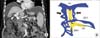
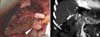
A 63-year-old female patient with hepatitis B-related liver cirrhosis was transferred to Daegu Catholic University Medical Center because of refractory ascites and hepatic encephalopathy. Preoperative Doppler ultrasonography and multidimensional computed tomography (MDCT) revealed the diffuse PVT that extended to the superior mesenteric vein (SMV) and splenic vein. In addition, marked dilated paracholedochal veins around the PV were being fed from the gastroepiploic veins and pancreaticoduodenal veins (Fig. 1A). Mesenteric venography demonstrated several findings, including complete invisibility of the main PV and SMV and large tortuous paracholedochal veins that extended to the proximal SMV (Fig. 1B). The donor was her 59-year-old brother. The estimated graft volume of the donor's right lobe as calculated by CT volumetry was 798 mL, 63.7% of the whole liver and the estimated graft-to-recipient weight ratio (GRWR) was 1.42%. Therefore, LDLT using a modified right lobe graft from her younger brother was performed. After complete mobilization of the recipient's liver, hepatic hilar dissection was attempted. However, dissection of the vascular pedicle at the hilum was too difficult due to severe fibrosis and a complex network of tortuous paracholedochal varices. Thus, after dissection and ligation of the hepatic arteries were performed, the PV and bile duct were clamped and transected en masse. Dissection of the native PV revealed that it had no obvious lumen and was replaced by a fibrotic, thrombosed cord. We identified two collateral veins with large orifices around the bile duct and transformed these large dilated collaterals into a common orifice with a venoplasty at their confluence. Several small collaterals which were not suitable for portal inflow were ligated. After declamping of the hilum, we ensured that the blood flow was adequate for portal inflow by spurting through the common orifice. We carried out usual end-to-end anastomosis of the donor PV to the large common orifice formed by the confluence of the markedly dilated paracholedochal veins using a running suture of 6-0 prolene (Fig. 2A). The graft was reperfused after completion of the PV anastomosis followed by hepatic artery reconstruction using a microsurgical technique. Roux-en-Y hepaticojejunostomy was performed because of severe periductal fibrosis and a narrowed caliber that could not maintain bile flow for duct-to-duct anastomosis. Intraoperative Doppler assessment showed adequate portal blood flow to the graft, and the PV patency was demonstrated by regular Doppler monitoring and MDCT (Fig. 2B).
The patient was discharged 3 weeks postoperatively without any complications and has been doing well for 22 months with normal graft function.
Case 2
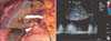
A 51-year-old man was admitted with massive ascites and general weakness due to alcoholic liver cirrhosis. Preoperative Doppler ultrasonography and MDCT demonstrated several dilated paracholedochal veins along hilum and diffuse PVT extended to SMV like the first case. The donor was his 30-year-old son. The preoperative CT volumetry demonstrated the estimated right to left lobe volumes to be 966 mL and 500 mL (65.9% and 34.1% in volume ratio) and the estimated GRWR was 1.19%. Thus, we planned to perform LDLT using a modified right lobe graft from his son. During the operation, we found chronic organized PVT with dilated paracholedochal collaterals along porta hepatis. The transected PV showed no venous flow; it contained chronic totally organized thrombi with fibrosis. Even though we tried to eversion thrombectomy, it was failed to obtain sufficient portal inflow to the graft. Unfortunately, the caliber of these collaterals was smaller than that of case 1 and venous flow through common orifice with venoplasty from their confluence was not sufficient for portal inflow. Because another approach to gain splanchnic inflow could not be available, we performed unification venoplasty with common colaca from paracholedochal veins and partially recanalized naitive PV to supplement unsatisfactory inflow through paracholedochal collaterals. This complex common orifice was anastomosed to the donor PV in end-to-end fashion as a single anastomosis (Fig. 3A). Hepatic artery and bile duct reconstruction was performed by the same method as case 1.
Intraoperative Doppler ultrasonography showed satisfactory portal blood flow in the liver graft and the good PV patency was demonstrated by serial postoperative Doppler studies(Fig. 3B) and MDCT. He was discharged without any serious complication and has remained stable without ascites, variceal bleeding for 21 months after transplantation.
DISCUSSION
In the early period of liver transplantation, PVT was considered to be a contraindication for the procedure because of the technical difficulties it entailed, especially the inability to gain an adequate portal supply [6]. However, several surgical techniques have been developed for deceased donor liver transplantation (DDLT), such as thrombectomy, interposition or a jump graft between the donor and recipient PV, cavoportal hemitransposition (CPHT), renoportal anastomosis (RPA), and PV arterializations [1,2]. With these surgical innovations, the results of DDLT for patients with PVT have been comparable to those of patients without PVT [1,6]. In spite of recent reports that described excellent outcomes in LDLT, several difficulties are associated with performance of these surgical innovations for pre-existing PVT in the setting of LDLT, such as the necessity of distal dissection of the vascular pedicle of the hilum, restricted availability of deceased donor vein grafts, and ethical concerns of the living donor [7]. Moreover, LDLT is still considered a surgically demanding challenge in the case of an extended PVT including the SMV with CTPV because this procedure is associated with perioperative morbidity and mortality as a result of rethrombosis and increased blood loss [8].
In 1986, Hiatt et al. [9] reported the first successful DDLT using a thin-walled bile duct varix as the inflow to the donor PV, and various alternatives were subsequently reported to overcome PVT with CTPV, such as CPHT, RPA, and portal arterializations [2,3]. In the present case, we planned to perform PV reconstruction using large dilated paracholedochal veins because it was demonstrated that these collaterals could provide the adequate portal inflow to the graft in preoperative MDCT and mesenteric venography. This technique offers some advantages over other surgical options using systemic blood flow as portal inflow. The advantages include the followings: (1) In PV reconstruction such as RPA, CPHT causes loss of hepatotrophic factors provided by PV inflow, which is believed to be essential for hepatic regeneration [2]. (2) In patients undergoing RPA and CPHT, persistent portal hypertension has been described with ascites, renal dysfunction, variceal bleeding, and hepatic encephalopathy [2,3]. (3) The long-term effects of PV inflow with systemic venous flow remain unknown [2]. Finally, (4) less vascular dissection is required and the operating time is decreased to a jump graft from the SMV to PV of the graft [10]. However, this technique, as mentioned above, also has disadvantages; e.g., using collateral veins to reconstruct the portal flow may increase the incidence of vascular complications such as thrombosis [1,10].
 XML Download