

 PDF
PDF ePub
ePub Citation
 Print
Print


INTRODUCTION
Deep vein thrombosis (DVT) is a serious medical problem that can result in complications such as pulmonary embolism, pulmonary hypertension, and chronic venous insufficiency of the lower extremities. Additionally, DVT represents a growing worldwide health concern, with an annual incidence of 48-95 events per 100,000 individuals in Caucasian populations [1,2,3]. The incidence, risk factors, and preventive methods for the development of DVT after general surgical procedures have been extensively documented in the literature [4,5]. However, studies dealing with DVT in kidney transplant recipients (KTRs) are scarce, and the data are contradictory. In fact, the true frequency and adequate preventive modalities of DVT in KTRs are undefined [6,7,8,9]. The 8th American College of Chest Physicians (ACCP) guidelines categorize KTRs as a moderate risk group and recommend routine thromboprophylaxis including anticoagulation [4]. The approximate risk of DVT in the group without thromboprophylaxis was reported as 10%-40%. However, these numbers are largely based on data from Caucasian individuals and may not be applicable to Korean KTRs because epidemiologic studies have shown that annual DVT incidence rates are significantly lower in Korean populations compared with Caucasian populations [10]. The aim of this study was to evaluate the incidence of lower extremity DVT during the first postoperative month in Korean patients who have undergone kidney transplantation (KT) and have received only mechanical thromboprophylaxis without chemoprophylaxis after KT.
METHODS
Approval for the study was obtained from the Institutional Review Board (No. KC12RISI0583).
Patients and study design
Beginning in November 2009, we performed routine serial postoperative color duplex ultrasonography (CDU) examinations and preoperative thrombophilic factor assays to identify the incidence and risk factors of lower extremity DVT in KTRs. Various demographic and clinical characteristics known to affect the development of DVT were also recorded prospectively on separate case report forms. From November 2009 to October 2011, we performed 228 KTs in our center. Among them, 41 patients were excluded due to failure to complete the postoperative CDU follow-up program. We analyzed 187 KTRs to estimate the incidence of lower extremity DVT and to identify its risk factors. We used only mechanical thromboprophylaxis with a graduated compression stocking (November 2009 to December 2010) or intermittent pneumatic compression (January 2011 to present) from the time of entry into the operation room until discharge. Recipients were usually discharged on postoperative day 14 and were followed for 1-month biweekly to detect clinical signs and symptoms of DVT.
Demographic, clinical, and laboratory data
To identify the risk factors of lower extremity DVT, various demographic, clinical, and laboratory characteristics were collected including donor and recipient age at time of KT, sex of donor and recipient, cause of end-stage renal disease, type and duration of renal replacement therapy, donor type, number of human leukocyte antigen mismatches, immunosuppressive medication (cyclosporine Avs. tacrolimus), preoperative comorbidities (diabetes mellitus and hypertension), acute rejection episodes within postoperative weeks 4, body mass index, history of DVT, history of antiplatelet therapy, history of erythropoietin therapy, platelet count, acute cytomegalovirus infection within postoperative weeks 4 (diagnosed by real-time quantitative polymerase chain reaction), and eight thrombophilic factors. Blood samples were collected before transplantation to detect the presence of thrombophilia. Eight thrombophilic factors, including factor V Leiden (G1691A) mutation, prothrombin (G20210A) mutation, antithrombin III, protein C activity, protein S activity, immunoglobulin G antiphospholipid antibody, lupus anticoagulant, and hyperhomocysteinemia, were included in our assay. And operation-related factors, including operative time (minute), amounts of intraoperative packed red blood cell transfusion (unit), duration of postoperative immobilization (day), duration (day) and total amounts (mL) of Jackson Pratt (JP) drains, and presence of lymphocele after operation were also reviewed.
Immunosuppression
The immunosuppressive protocol in this study period has been described in detail elsewhere [11]. In summary, 32 high-risk KTRs (20 ABO incompatible and 12 positivecrossmatch patients) received sequential quadruple immunosuppressive therapy consisting of basiliximab (Simulect, Novartis Pharmaceuticals, Basel, Switzerland), tacrolimus (Tacrobell, Chong Kun Dang Pharm, Seoul, Korea, or Prograf, Astellas, Toyama, Japan), corticosteroids, and either mycophenolatemofetil (Cellcept, Roche, Nutley, NJ, USA) or mycophenolate sodium (Myfortic, Norvatis Pharmaceuticals). These patients also received a single dose of rituximab (Roche Pharm, Renach, Switzerland) and plasma exchange on an every other day schedule followed by intravenous immunoglobulin. Conventional KTRs (155 patients) received induction therapy with basiliximab and maintenance therapy with tacrolimus or cyclosporine A (Neoral, Novartis Pharmaceuticals), mycophenolatemofetil or mycophenolate sodium, and corticosteroids.
Operation
Recipient surgery was performed through an extraperitoneal "hockey stick" incision with creation of standard vascular anastomoses and extravesical ureteroneocystostomy. JP drains (Cardinal Health, McGaw Park, IL, USA) remained in the extraperitoneal space until the drainage was less than 50 mL/day for 2 consecutive days.
Diagnosis of DVT
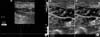
DVT was investigated and diagnosed by CDU (Philips HD11XE System, Markham, ON, Canada). In the present study, 187 patients who had undergone KT had bilateral, whole-leg CDU on postoperative week 1 (day 7 ± 2 days), week 2 (day 14 ± 2 days), and week 4 (day 28 ± 3 days), or earlier if clinically indicated, in order to detect lower extremity DVT. The deep venous system of both lower extremities was extensively evaluated using the compression technique of Cronan et al. [12] supplemented with duplex ultrasonography and color Doppler studies (Fig. 1).
Statistics
All data are presented as mean ± standard deviation. Comparisons between groups were performed using the chi-square test or Student t-test. A P-value < 0.05 was considered statistically significant. All statistical analyses were carried out with the SAS 9.1 (SAS Institute Inc., Cary, NC, USA).
RESULTS
Incidence and clinical characteristics of DVT during the first 4 posttransplantation weeks
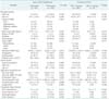
Of the 187 KTRs, four patients (2.1%) developed an acute lower extremity DVT within 1 month of KT. Several features of these patients are given in Table 1. Only one of the four had clinical evidence of a DVT while the other three were asymptomatic and diagnosed during scheduled CDU. In terms of DVT location, three developed in the same side of graft, one developed in bilaterally. The median time for DVT development was 14 days (range, 2-28 days) after KT and only one patient experienced a DVT within postoperative week 1. All DVTs developed in the graduated elastic stocking (GES) group compared with the pneumatic compression device (PCD) group, however this finding was not statistically significant (P = 0.058).
Comparison of demographic, clinical, and laboratory factors of KTRs with and without DVT in the lower extremities
Demographic, clinical, and laboratory factors of the two groups are shown in Table 2. A total of 22 different factors were analyzed to determine the risk factors for DVT. There were no statistically significant differences between patients with DVT and those without. There were no statistically significant differences between the GES and PCD groups.
The comparison of thrombophilic factors of KTRs with and without lower extremity DVT
Among the 187 KTRs, 160 had eight factors tested, 23 patients had seven factors tested, and only one recipient had fewer than six factors tested before transplantation (Table 3). The most common hypercoagulable abnormality was hyperhomocysteinemia (114/178 patients, 64%). Cases of factor V Leiden mutation or prothrombin gene 20210A mutation were not identified. There were 151 patients with ≥1 thrombophilic factor and positivity of tested thrombophilic factors was17.1% patients (249 of 1,458). In the comparison of patients who had developed DVT and those who had not, there were no differences in terms of individual thrombophilic factor, number of patients with more than 1 thrombophilic factor, or the percentage of positivity of tested thrombophilic factors (Table 3).
DISCUSSION
According to the 8th ACCP guidelines, KTRs are categorized as a moderate risk group for DVT and the approximate risk of DVT without thromboprophylaxis was reported to be 10%-40% [4]. When the "Caprini Risk Assessment Model" [13] was applied to our patients, most recipients (95.7%, 179/187) were categorized at the highest risk for DVT. Incidence of DVT in this highest risk group without thromboprophylaxis is expected to be 40%-80%. However, the incidence of DVT after KT in this study was only 2.1% (4/187). This figure is lower than the 9%-20% quoted after general surgical procedures of similar magnitude without prophylaxis [14]. It is also lower than the 16.1%-57.1% quoted in other prospective studies of DVT in Caucasian KTRs [6,15,16].
There are two possible explanations for the lower incidence rates of DVT in our patients even without thromboprophylaxis: (1) difference in the prevalence of thrombophilic factors in a Korean population; and (2) the use of routine closed suction drainage. First, a difference in the incidence of DVT between ethnicities has been reported [17] and several reports suggest a low prevalence of DVT in Asian populations [18,19,20]. Factors associated with this difference in an Asian population are most likely reflected by a different prevalence of thrombophilic factors between Asian people and Caucasian people. There are also data supporting genetic differences as a partial explanation of the lower risk of DVT in Asian people. Factor V Leiden mutations and the prothrombin G20210A mutations are the most potent inherited thrombophilic factors and are highly prevalent in Caucasian people, but are almost nonexistent in Asian people [21]. The prevalence of factor V Leiden and prothrombin G20210A gene mutations is 4.8%-11.5% and 4%-6%, respectively, in Caucasian people, and are both rarely reported in Asian populations [21,22,23,24]. In our patient population, factor V Leiden mutations and prothrombin G20210A mutations were absent in all tested patients.
Second, a lymphocele is a common and well-known complication that occurs in 1% to 26% of KTRs and can cause DVT after compression of the external iliac vein [25]. Derweesh et al. [26] studied the effect of prophylactic drain placement on the incidence of lymphocele and DVT in KTRs. They found that placing a drain during the operation significantly decreased the incidence of symptomatic lymphoceles (19.0% vs. 2.5%) and DVT (14.3% vs. 4.9%) [26]. We also routinely used prophylactic drains and none of the study patients experienced a symptomatic lymphocele. These observations suggest that prevention of lymphocele formation with a routine drain is one of the reasons for the low incidence of DVT in our study.
To date, there has been no consensus regarding the optimum type of thromboprophylaxis in KTRs. Patients with renal failure are physiologically different from the average surgical patient because uremia increases the risk of bleeding. Therefore, the use of prophylactic anticoagulation has to be well balanced with the risk of postoperative bleeding. Some authors reported higher incidence of bleeding complications associated with routine use of prophylactic anticoagulation for KTRs [27,28] and several reports have suggested a low prevalence of DVT and pulmonary embolism in Asian populations, particularly in the Far East, when compared to western populations [18,19,20]. For these reasons, we used mechanical thromboprophylaxis modalities instead of anticoagulation as thromboprophylaxis and experienced a fairly low incidence of DVT. In our experience, PCD seems to be more effective at preventing lower extremity DVT in KTRs compared with GES.
This study has several limitations. Although a retrospective study of DVT such as this is prone to underestimate its true incidence, we performed a serial CDU according to a specific schedule and results were systematically recorded in this study. Second, CDU was used to detect postoperative DVT in this study. Venography is still considered the gold standard for the detection of DVT; however, venography is invasive, may induce nephrotoxicity, and is time-consuming and difficult to perform repeatedly. In contrast, DUS can be performed safely without any serious complications and our registered vascular technologists experienced and was able to detects mall thrombi in asymptomatic patients.
In conclusion, this is the first report concerning the incidence of DVT in Asian KTRs. The incidence of DVT without chemoprophylaxis in this study was only 2.1%. This figure is lower than that of Caucasian KTRs. This study suggests that different combinations of genetic thrombophilic polymorphisms with use of a routine JP drain led to the lower prevalence of DVT after KT in the current series of patients who did not receive DVT chemoprophylaxis. Thus, we do not recommend routine use of chemoprophylaxis to prevent DVT for Korean KTRs. However, we do suggest further randomized controlled trials to compare the efficacy and adverse effects between chemoprophylaxis and mechanical thromboprophylaxis in this particular patient population.


 XML Download