

 PDF
PDF ePub
ePub Citation
 Print
Print


INTRODUCTION
Internal biliary fistula (IBF) is an abnormal passage between the biliary system and visceral organ. Three kinds of IBF including biliobiliary, bilioenteric, and bronchobiliary have been reported in the literature. The bilioenteric type are rare and it's most common findings in radiology are pneumobilia. However, preoperative diagnosis is usually quite difficult due to both ambiguous symptoms and radiologic findings [1].
We herein describe our experience with an incidental cholecystojejunal fistula in laparoscopic cholecystectomy. Preoperative diagnosis is an emphysematous cholecystitis and the patient did not have any other combined diseases and symptoms. To our knowledge, this is the first report on incidental cholecystojejunal fistula.
CASE REPORT
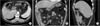
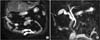
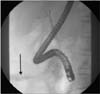
A 61-year-old woman with several years' history of intermittent epigastric pain was admitted to Soonchunhyang University Cheonan Hospital with right upper abdominal pain and myalgia which were aggravated during the first day. She had no medical and operation history except hypertension. Physical examination revealed rigid abdomen with tenderness and mild rebound tenderness on the right upper abdomen. Also, she had a fever at 38.0℃. Other symptoms were insignificant. Laboratory findings showed no leukocytosis, but CRP was elevated to 139, while liver function test and tumor marker including CEA, CA 19-9 and α-FP were within the normal limits. Abdominal ultrasound showed gallbladder (GB) stones, common bile duct (CBD) dilatation with CBD stones. Abdominal CT scan (Fig. 1), and MR cholangiopancreatography (MRCP) (Fig. 2) revealed the presence of GB stones with minimal wall enhancement on postcontrast CT and the pneumobilia in GB. However, as the CBD stones were not clear, we performed endoscopic retrograde cholangiopancreatography (ERCP) (Fig. 3) and endoscopic sphincterotomy. However, there were no CBD stones.
Consequently, laparoscopic cholecystectomy was carried out. Operative findings revealed that the fundus of GB was connected with proximal jejunum. So we performed laparoscopic cholecystectomy and fistulectomy with jejunal partial resection by Endo-GIA (Covidien plc, Dublin, Ireland) stapling device. On the pathologic reports, chronic cholecystitis with cholelithiasis and cholecystojejunal fistula with chronic inflammation was found. The patient was discharged with an uneventful recovery on the fourth postoperative day.
DISCUSSION
The cause of IBF is both adhesion and erosion between the biliary tract and the visceral organ due to the inflammatory condition from cholelithiasis, peptic ulcer, malignancy, and Crohn's disease. Especially, chronic cholecystitis with GB stones is considered in about 75% of major etiology. Our case is also related with chronic cholecystitis. Although the inflammation of the image findings was not severe, she had a complaint of recurrent epigastric pain for several years. On the pathologic reports, chronic inflammation was also found in cholecystojejunal fistula.
Cholecystoenteric fistula (CF) is a bilioenteric type of IBF, and it's incidence is reported to be between 0.15% to 5% of the biliary disease. It's locations, in order of frequency, have been reported duodenum, colon and stomach [2]. The most common type of CF is cholecystoduodenal fistula which is reported to be about 80% of CF cases. The cholecystocolonic type is reported about 10%. And then, cholecystogastric fistula is reported [1]. However, to our knowledge, cholecystojejunal fistula has been reported in only one case which is related to hepatocellular carcinoma [3].
The preoperative radiologic diagnosis is very difficult as only remarkable findings are considered pneumobilia. However, as it can also occur due to other causes, it is nonspecific. Laboratory findings are also unremarkable for differential diagnosis. In our case, although the patient has complained epigastric discomfort for several years, the CT scans showed no abnormal findings except for the pneumobilia in GB. MRCP and ERCP also revealed no other abnormal findings such as GB stones, CBD stones, and malignancy. But after the operation, we reviewed the image findings and found that the GB and the small bowel are adherent with each other.
It has been known that the standard treatments are cholecystectomy and repair of fistula opening. Although s everal cases are reported about laparoscopic management, there are reported high conversion rate due to CBD exploration and T-tube insertion [4]. In addition, the other cause of high conversion rate is complete resection of the enteric lesion. In order to perform differential diagnosis with malignancy and prevent the enteral perforation from ischemic change of remnant enteric fistula, laparoscopic procedure is not feasible [5].
However, recently, laparoscopic skills of a surgeon including laparoscopic duodenal mobilization, laparoscopic suture and knotting have been improved. The ligation is also easier thanks to the surgical instruments such as Endo-loop and Endo-GIA stapling devices. Hence, the open conversion rate has been decreased, and as a consequence, reports about laparoscopic management of CF have been increased, it is not contraindication anymore [4]. In our case, small bowel wedge resection was also performed by Endo-GIA stapling device, and the patient was discharged without an event.
In conclusion, this is the first report on incidental cholecystojejunal fistula not combined with any other disease and was treated with laparoscopic procedure. It was misdiagnosed as emphysematous cholecystitis due to pneumobilia in GB. Thus, a careful diagnosis of radiologic findings is essential. And laparoscopic management of IBF is no longer a contraindication. Laparoscopic fistulectomy and enteral wedge resection by skilled laparoscopic surgeon is both safe and feasible.
 XML Download