Abstract
Purpose
This study was performed to analyze the surgical pathology results of the "atypia of undetermined significance" (AUS) category from thyroid fine needle aspiration (FNA) and to describe the characteristics to distinguish a malignant from a benign nodule.
Methods
A retrospective analysis was done on 116 patients who underwent thyroid surgery from December 2008 to December 2012, following a diagnosis of AUS from preoperative thyroid FNA. We investigated the age, gender, size and site of the nodules, ultrasonographic criteria, cytological features, the number of atypia results after repeated FNAs, surgical method, and final pathologic results.
Results
Sixty-five out of 116 patients underwent total thyroidectomy and the rest had partial thyroidectomy. The final pathologic results were 41 malignancies (35.3%) and 75 benign diseases (64.7%). AUS was divided into group 1: 'cannot rule out malignancy' or group 2: 'cannot rule out follicular neoplasm'. After surgery, group 1 revealed papillary thyroid cancer in most cases and group 2 revealed follicular adenoma in most cases. Age over 40 years, ultrasonographic findings suggestive of malignancy, more than 2 results of atypia from repeated FNAs and nodules less than 2 centimeters were risk factors for malignancy on univariate analysis. Multivariate analysis showed that ultrasonographic findings suggestive of malignancy was a significant risk factor for malignancy.
Fine needle aspiration (FNA) of thyroid nodules is the initial diagnostic test, establishing whether these lesions are benign or malignant. Preoperative FNA in thyroid benign nodules reduced unnecessary surgery. According to reports, the sensitivity and specificity on FNA were 50%-90% [1-3]. However, cytology has limitations; accuracy is lower in atypia nodule and in follicular neoplasm [4,5]. In particular, the final pathology of atypical nodules on thyroid FNAs was malignant in 6%-48% of cases [6,7]. Other clinical factors should be considered because it is difficult to determine whether the atypia seen in cytology is benign or malignant. Clinical predictors (nodule size, sex, and age), immunohistochemical markers (HBME, galectin-3), molecular analysis (BRAF, PAX-8/PPAR rearrangement), ultrasonography (malignant criteria or elastography) and FDG-PET have been assessed as potential predictors of malignancy, but none of these were shown to be specific [8-11].
The Bethesda six-category diagnostic scheme consisted of benign, atypia of undetermined significance (AUS), suspicious for follicular neoplasm, suspicious for malignancy, malignant, and unsatisfactory [12]. An important aspect of this system is that each diagnostic category is associated with relative risk of malignancy.
This study was done to analyze the pathological results of the AUS category and to find out the risk factors of malignancy.
One hundred sixteen patients underwent thyroid surgery due to AUS from preoperative FNA between December 2008 and December 2012. The indications of surgery for AUS patients were suspicious findings on clinical or radiologic evaluation, repeated atypia results from FNA, patients' willing to undergo surgery due to the fear of malignancy or diagnostic confirmation. We retrospectively reviewed patients' medical records and other clinicopathologic data after obtaining approval from our Institutional Review Board. All patients underwent thyroid function tests and ultrasonography for the initial staging work-up before their surgery and were followed-up with computed tomography as necessary. In all patients, ethanol-fixed Papanicolaou-stained direct smears were performed under ultrasound guidance, using 24-gauge needles. FNA was analyzed according to the Bethesda classification, and an AUS result is given when the cytologic findings are not convincingly benign, yet the degree of cellular or architectural atypia is not sufficient for an interpretation of "follicular neoplasm" or "suspicious for malignancy" [13]. Patients were excluded from the study if they had follow-up FNA with the diagnosis of follicular neoplasm or suspicious for malignancy that did require further surgery.
Clinicopathologic data included age, sex, the size and site of the nodules, ultrasonographic criteria, cytological features, the number of atypia results after repeated FNAs, surgical method and final pathologic results. Ultrasonographic criteria for malignancy include any of following findings; hypoechogenecity, irregular border, calcification, increasing size, and abnormality of vasculature.
AUS was divided into two groups as follows: 'cannot rule out malignancy' (group 1) or 'cannot rule out follicular neoplasm' (group 2). The cytology of AUS group 1 has a minor population of atypical cells showing nuclear enlargement, pleomorphism, abnormal chromatin and prominent nucleoli in a benign-appearing sample (especially in patients with chronic thyroiditis or adenomatous goiter), but the degree of atypia is insufficient for the general category of "suspicious for malignancy". The AUS group 2 shows prominent population of microfollicles in a sparsely cellular aspirate with scant colloid that does not otherwise fulfill the criteria for "follicular neoplasm/suspicious for follicular neoplasm".
Statistical significance between subgroups was determined using chi-square test and Student t-test. A P-value less than 0.05 was considered significant. Statistical analysis was performed using PASW ver. 18.0 (SPSS Inc., Chicago, IL, USA). Logistic regression analysis was performed to evaluate the risk factors for the presence of malignancy.
The mean age of the study population was 46 years, ranging from 14 to 76 years old. The study group consisted of 91 females and 25 males (3.6:1). Total thyroidectomy was performed in 65 of the 116 patients and partial thyroidectomy in remaining 51 patients. Among the 51 patients who had partial thyroidectomy, 15 had a malignant tumor and completion thyroidectomy was later performed in 6 patients. The remaining 9 patients had no recurrences upon follow-up (Fig. 1).
Among the 116 cases, 41 (35.3%) had final diagnosis of malignancy and 75 (64.7%) had benign lesions. Of the 41 patients who had histologically confirmed thyroid cancer, 25 were papillary carcinoma, 8 had follicular variant of papillary carcinoma, 5 had follicular carcinoma, 2 had medullary carcinoma, and 1 was poorly differentiated carcinoma. Of the 75 benign lesions, nodular hyperplasia was present in 40 patients, 30 had follicular adenoma, 2 had Hürthle cell adenoma, 2 had Hashimoto's thyroiditis, and 1 had Riedel's thyroiditis. AUS was divided into 'cannot rule out malignancy' (group 1) or 'cannot rule out follicular neoplasm' (group 2) (Table 1). The most common malignant pathology in group 1 was papillary thyroid cancer (36.4%), whereas the most common benign pathology was nodular hyperplasia (34.5%). In group 2, the most common malignant pathology was papillary thyroid cancer (8.2%) and follicular variant papillary thyroid cancer (8.2%), whereas the most common benign pathology was follicular adenoma (37.7%).
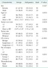
A comparison of age, sex, tumor size, site, sonographic criteria, and results of final histology from patients with benign or malignant lesion are detailed in Table 2. Univariate analysis showed no differences between the benign and malignant groups by sex, tumor site, and operation type. The average age of patients with malignant tumors (49.8 ± 13.2) was older than in those with benign diseases (43.4 ± 15.1) (P = 0.023). The malignant nodules (mean size, 1.72 ± 1.22 cm) were smaller than the benign nodules (mean size, 2.47 ± 1.47 cm) (P = 0.006). The malignancy rate was 5.3% in patients without sonographic criteria for malignancy. If one of the sonographic malignant features is present, the risk of malignancy is 40.4%; if two are present, the risk of malignancy is 66.7% (P < 0.005, likelihood ratio test). The risk of malignancy was higher in patients with more than 2 results of atypia from repeated FNAs (51.9%, 14 of 27 patients) in comparison with patients with only 1 result of atypia from repeated FNAs (30.3%, 27 of 89 patients) (P = 0.041). There were significantly more malignant tumors among patients in group 1 than in group 2 (47.3% vs. 24.6%, P = 0.011).
Age over 40 years, ultrasonographic findings suggestive of malignancy, more than 2 results of atypia from repeated FNAs and nodules less than 2 centimeters were statistically significant risk factors of malignancy on univariate analysis. Multivariate analysis showed that ultrasonographic findings suggestive of malignancy was a significant risk factor of malignancy (Table 3).
FNA is an accurate method for diagnosing a thyroid nodule. FNA has a sensitivity of around 61.8%-98.4% and a specificity of approximately 71.4%-100% [3,14,15]. However, the predictive value of atypia on cytology for a malignancy is not known. Previous studies found that 20% of AUS presenting in the thyroid nodule were diagnosed using FNA and 15% to 47% of these thyroid nodules cases were later histologically proved to have a malignancy [16-18].
Atypia in the thyroid presents clinicians with a diagnostic dilemma. Several clinicians have closely examined their patients presenting with atypia in the thyroid in an attempt to elucidate some clinical features that might determine whether patients should be observed or should undergo surgery [19]. This study analyzed the pathological results of 116 patients who underwent thyroid surgery due to AUS from preoperative FNA to define the characteristics that may help in distinguishing malignant from benign nodules.
In this study, among the 116 patients, 41 (35.3%) had malignant tumor. This malignancy rate is similar with that of follicular neoplasm rather than atypia category. The reason may be that clinicians select patients for surgery by considering the cytologic findings as well as other clinical factors. According to our results, papillary carcinoma is the most common malignant type (n = 25) after surgery. Some authors found that papillary carcinoma and follicular neoplasm rates were similar, or that follicular variant of papillary carcinoma was the most common type [10,20,21].
Male gender was associated with malignancy by some studies [8,9] but other studies reported no significant difference in the rate of malignancy between the sexes [10,11,22]. We found no significant difference in the incidence of malignant thyroid tumor between male and female patients. Some reports have suggested that a significant predictor of malignancy was the age of patient [8,18,23,24]. However, other studies showed that age was not associated with an increased risk of malignancy [10,11,21]. In the present study, older age (≥40 years) is associated with an increased risk of malignancy. But no significant difference was seen after multivariate analysis. This result may be due to patient selection bias that surgeons consider age as a risk of malignancy when they decide the surgery.
In some studies, the risk of malignancy has been shown to increase with nodule size. Sippel et al. [21] showed that tumor size was associated with an increased risk of malignancy but McHenry at al. [25] found no significant difference in size between benign and malignant thyroid nodules. We grouped the thyroid nodules into size categories in 2 cm increments and malignancy was more frequently found in patients with a nodule <2 cm than in patients with a nodule ≥2 cm (44.3% vs. 25.5%, P = 0.034).
According to the Bethesda classification, AUS is defined as a category in which the cytomorphological findings are not representative of benign lesion such as a hyperplastic/adenomatoid nodule, yet the degree of cellular or architectural atypia is not sufficient for an interpretation of "follicular neoplasm" or "suspicious for malignancy". The risk for malignancy in this category is approximately 5%-15% [12]. In the present study, there were more malignant tumors among patients in group 1 than in group 2 (47.3% vs. 24.6%, P = 0.011). We did not analyze factors associated with the pathologist, although results of FNA are pathologist dependent. Therefore, there is no significant differences compared with the findings of ultrasound (Table 3).
According to the NCI conference guideline, atypia categories can benefit from repeat aspiration or correlation with clinical and radiological findings [12]. Nayar and Ivanovic [26] have published their experience with the above guideline. Among follicular lesions, follow-up FNA was performed in 31%, of which 58% had surgery and 12% were malignant. This is similar to the outcome of follicular neoplasm, supporting that surgery is justified in this subgroup. In our current study, among those with repeat AUS diagnosis on follow-up FNA, of which 27 had surgery and 14 (51.9%) were diagnosed with malignancy (Table 2).
Radiological correlation may be helpful in improving the positive predictive value of the AUS category. A previous study reported that the sonographic findings of malignant thyroid nodule include hypoechogenicity, irregular borders, calcifications, abnormalities of vasculature and increasing size [3]. In this study, the malignancy rate was 5.3% in patients without sonographic criteria for malignancy. If one of the sonographic malignant feature is present, the risk of malignancy is 40.4%; if two are present, the risk of malignancy is 66.7% (P < 0.005, likelihood ratio test). The presence of sonographic malignant features have been shown to be associated with an increased risk of malignancy on multivariate analysis.
On the basis of our study, the risk of malignancy is 35.3% among cases with AUS on thyroid FNA. However, if the ultrasonographic findings did not suggest malignancy, the risk of malignancy is 5.3%. From the results of our study, we recommend that ultrasonographic criteria should be considered along with other clinicopathological findings such as age, nodule size, number of atypia, cytologic features for evaluation of the risk for malignancy in thyroid AUS nodule.
Figures and Tables
Table 1
Result of pathologic diagnosis according to fine needle aspiration cytology groups

Group 1, cannot rule out malignancy; Group 2, cannot rule out follicular neoplasm; FA, follicular adenoma; FTC, follicular thyroid cancer; HA, Hürthle cell adenoma; HT, Hashimoto's thyroiditis; MTC, medullary thyroid cancer; NH, nodular hyperplasia; PDTC, poorly differentiated thyroid cancer; PTC, Papillary thyroid cancer; PTCFV, follicular variant papillary thyroid cancer; RT, Riedel's thyroiditis.
References
1. Baloch ZW, Sack MJ, Yu GH, Livolsi VA, Gupta PK. Fine-needle aspiration of thyroid: an institutional experience. Thyroid. 1998; 8:565–569.
2. Gharib H, Goellner JR, Johnson DA. Fine-needle aspiration cytology of the thyroid: a 12-year experience with 11,000 biopsies. Clin Lab Med. 1993; 13:699–709.
3. Gharib H, Goellner JR. Fine-needle aspiration biopsy of the thyroid: an appraisal. Ann Intern Med. 1993; 118:282–289.
4. Alexander EK. Approach to the patient with a cytologically indeterminate thyroid nodule. J Clin Endocrinol Metab. 2008; 93:4175–4182.
5. Baloch ZW, Cibas ES, Clark DP, Layfield LJ, Ljung BM, Pitman MB, et al. The National Cancer Institute Thyroid fine needle aspiration state of the science conference: a summation. Cytojournal. 2008; 5:6.
6. Ohori NP, Schoedel KE. Variability in the atypia of undetermined significance/follicular lesion of undetermined significance diagnosis in the Bethesda System for Reporting Thyroid Cytopathology: sources and recommendations. Acta Cytol. 2011; 55:492–498.
7. VanderLaan PA, Marqusee E, Krane JF. Clinical outcome for atypia of undetermined significance in thyroid fine-needle aspirations: should repeated fna be the preferred initial approach? Am J Clin Pathol. 2011; 135:770–775.
8. Baloch ZW, Fleisher S, LiVolsi VA, Gupta PK. Diagnosis of "follicular neoplasm": a gray zone in thyroid fine-needle aspiration cytology. Diagn Cytopathol. 2002; 26:41–44.
9. Tuttle RM, Lemar H, Burch HB. Clinical features associated with an increased risk of thyroid malignancy in patients with follicular neoplasia by fine-needle aspiration. Thyroid. 1998; 8:377–383.
10. Rago T, Di Coscio G, Basolo F, Scutari M, Elisei R, Berti P, et al. Combined clinical, thyroid ultrasound and cytological features help to predict thyroid malignancy in follicular and Hupsilonrthle cell thyroid lesions: results from a series of 505 consecutive patients. Clin Endocrinol (Oxf). 2007; 66:13–20.
11. Wiseman SM, Baliski C, Irvine R, Anderson D, Wilkins G, Filipenko D, et al. Hemithyroidectomy: the optimal initial surgical approach for individuals undergoing surgery for a cytological diagnosis of follicular neoplasm. Ann Surg Oncol. 2006; 13:425–432.
12. Baloch ZW, LiVolsi VA, Asa SL, Rosai J, Merino MJ, Randolph G, et al. Diagnostic terminology and morphologic criteria for cytologic diagnosis of thyroid lesions: a synopsis of the National Cancer Institute Thyroid Fine-Needle Aspiration State of the Science Conference. Diagn Cytopathol. 2008; 36:425–437.
13. Layfield LJ, Cibas ES, Baloch Z. Thyroid fine needle aspiration cytology: a review of the National Cancer Institute state of the science symposium. Cytopathology. 2010; 21:75–85.
14. Kim YM, Kim TC, Moon YB, Rho YS, Park YM. The clinical significance of fine needle aspiration cytology in the surgical management of thyroid nodules. Korean J Otolaryngol-Head Neck Surg. 1995; 38:1081–1087.
15. Agrawal S. Diagnostic accuracy and role of fine needle aspiration cytology in management of thyroid nodules. J Surg Oncol. 1995; 58:168–172.
16. Hamburger JI. Diagnosis of thyroid nodules by fine needle biopsy: use and abuse. J Clin Endocrinol Metab. 1994; 79:335–339.
17. Bahar G, Braslavsky D, Shpitzer T, Feinmesser R, Avidan S, Popovtzer A, et al. The cytological and clinical value of the thyroid "follicular lesion". Am J Otolaryngol. 2003; 24:217–220.
18. Kim ES, Nam-Goong IS, Gong G, Hong SJ, Kim WB, Shong YK. Postoperative findings and risk for malignancy in thyroid nodules with cytological diagnosis of the so-called "follicular neoplasm". Korean J Intern Med. 2003; 18:94–97.
19. Hegedüs L. Clinical practice: the thyroid nodule. N Engl J Med. 2004; 351:1764–1771.
20. Tysome JR, Chandra A, Chang F, Puwanarajah P, Elliott M, Caroll P, et al. Improving prediction of malignancy of cytologically indeterminate thyroid nodules. Br J Surg. 2009; 96:1400–1405.
21. Sippel RS, Elaraj DM, Khanafshar E, Kebebew E, Duh QY, Clark OH. Does the presence of additional thyroid nodules on ultrasound alter the risk of malignancy in patients with a follicular neoplasm of the thyroid? Surgery. 2007; 142:851–857.
22. McHenry CR, Thomas SR, Slusarczyk SJ, Khiyami A. Follicular or Hürthle cell neoplasm of the thyroid: can clinical factors be used to predict carcinoma and determine extent of thyroidectomy? Surgery. 1999; 126:798–802.
23. Schlinkert RT, van Heerden JA, Goellner JR, Gharib H, Smith SL, Rosales RF, et al. Factors that predict malignant thyroid lesions when fine-needle aspiration is "suspicious for follicular neoplasm". Mayo Clin Proc. 1997; 72:913–916.
24. Tyler DS, Winchester DJ, Caraway NP, Hickey RC, Evans DB. Indeterminate fine-needle aspiration biopsy of the thyroid: identification of subgroups at high risk for invasive carcinoma. Surgery. 1994; 116:1054–1060.
25. McHenry CR, Huh ES, Machekano RN. Is nodule size an independent predictor of thyroid malignancy? Surgery. 2008; 144:1062–1068.
26. Nayar R, Ivanovic M. The indeterminate thyroid fine-needle aspiration: experience from an academic center using terminology similar to that proposed in the 2007 National Cancer Institute Thyroid Fine Needle Aspiration State of the Science Conference. Cancer. 2009; 117:195–202.


 PDF
PDF ePub
ePub Citation
 Print
Print


 XML Download