

 ePub
ePub Citation
 Print
Print


INTRODUCTION
Gastric volvulus is defined as the rotation of the stomach or part of the stomach by more than 180°, which creates a closed loop obstruction [1]. It can lead to ulceration, perforation, hemorrhage, or ischemia of the incarcerated stomach. Quick detection and prompt surgical correction are the mainstays of therapy for acute gastric volvulus [2]. The traditional treatment of gastric volvulus includes laparotomy, gastric detorsion, and gastric fixation. If the volvulus is secondary to a diaphragmatic defect, the defect should be corrected [3]. Laparoscopic reduction and gastropexy have been used to treat chronic gastric volvulus [4,5]. The present report describes the case of a 50-year-old patient with acute primary gastric volvulus who was treated by laparoscopic reduction with percutaneous endoscopic gastrostomy (PEG). While this approach has been employed for chronic gastric volvulus, to our knowledge, it has not been used for the treatment of acute gastric volvulus [6].
CASE REPORT
A 50-year-old man with a medical history of subdural hematoma 8 years previously recently reported the sudden onset of pain in the epigastric area. He visited the local hospital and was checked by simple abdominal X-rays, abdominal computed tomography (CT) and gastroscopy. He was diagnosed with acute gastric volvulus and was transferred to our hospital for an emergency operation.
His vital signs were as follows: temperature, 36.9℃; heart rate, 70/min; blood pressure, 170/90 mmHg; and respiration, 16/min. On physical examination, the abdomen was found to be distended with upper abdominal tenderness and rebound tenderness. Laboratory values were within normal limits.
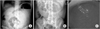
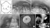
Fig. 1 shows the initial simple abdominal X-ray and the endoscopic image. Both the abdominal erect and supine X-ray views reveal the distention and air-fluid levels in the stomach (Fig. 1A, B). The endoscopic image shows the congested and edematous gastric wall (Fig. 1C). However, endoscopic reduction of the acute gastric volvulus could not be performed. Fig. 2A depicts the twisted axis: the stomach was rotated along the axis that joins the mid and lesser curvatures (mesentero-axial volvulus). The coronal abdominal CT view provides detailed information about the gastric volvulus (Fig. 2B, C). In Fig. 2B, arrow ① indicates the esophagus and gastroesophageal junction, and arrow ② shows that the body portion is superior to the fundus area. Arrow ③ in Fig. 2C shows that the gastric low body is located below the diaphragm and is connected to the duodenum.
The patient complained of severe abdominal and rebound tenderness. Since this is one of the symptoms and a sign of gastric strangulation, an emergency operation was scheduled.
Operative technique and postoperative course
After administering a general anesthetic, the patient was placed in the inverted Y position. The surgeon stood to the right of the patient and the first assistant stood opposite the surgeon. The camera operator stood between the legs of the patient. The umbilical camera port was established by using an open technique. After establishing a pneumoperitoneum, four ports were placed (two 11-mm and two 5-mm ports) (Fig. 3A). Intracorporeal CO2 pressure was maintained at 12 mmHg. The operation commenced with a survey of the intraabdominal cavity. Blood stains in the greater omentum were detected along with a mass-like twisted stomach below the diaphragm (Fig. 3B). The reduction of the gastric volvulus was performed by placing traction on the greater omentum with two atraumatic bowel graspers. Normal stomach anatomy was then observed (Fig. 3C). Blood and hematoma could be seen in the spleen hilum. Endoscopist placed the PEG in the midbody greater curvature side (Fig. 3D). An endoscopic image of the PEG after its insertion is shown in Fig. 3E. An upper gastrointestinal (GI) contrast study was performed on the third postoperative day and complete reduction of the stomach was confirmed without any evidence of obstruction (Fig. 3F). The patient was put on a diet on the fourth postoperative day and was discharged on the sixth.
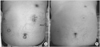
The patient visited the outpatient clinic on the fourteenth postoperative day. A photo of his abdomen is shown in Fig. 4A (the blue circles indicate the port sites). He had resumed his regular diet. He was readmitted for the removal of the PEG 2 months after the initial operation. After fasting beginning at midnight, the silicone PEG was removed by extracting the catheter. The patient fasted for 2 days and then resumed his regular diet. He was discharged after 3 days. Five months after his initial operation, the abdominal scars had almost completely disappeared (Fig. 4B).
DISCUSSION
Although rare, acute gastric volvulus is a GI condition that has catastrophic complications if it is not treated promptly. Early detection followed by surgical reduction is the key to successfully managing this disorder [2]. Although endoscopic reduction can be achieved in patients with chronic forms of gastric volvulus, it is not recommended for initial therapy in an acute setting, except for the treatment of high risk patients [3].
The first reported case of gastric volvulus was by Berti in 1866 after he performed an autopsy on a 60-year-old woman. Berg is credited with the first successful operative reduction of a gastric volvulus in 1896. In 1904, Borchardt described the classic triad of severe epigastric pain and distention, vomiting, followed by violent retching without the ability to vomit, and difficulty or inability to pass through a nasogastric tube. Gastric volvulus occurs when the stomach rotates along its longitudinal axis (organo-axial volvulus), or along the axis joining the midlesser and greater curvatures (mesentero-axial volvulus). One-third of gastric volvulus cases are caused by congenitally lax gastrocolic ligaments or are acquired. Two-thirds of gastric volvulus cases are secondary to paraesophageal hernia or a diaphragmatic defect [3].
The signs and symptoms of gastric volvulus depend upon the rapidity of onset, the degree of rotation, and the amount of obstruction [3]. Clinically, gastric volvulus may present either as an acute abdominal emergency or as a chronic recurrent problem. Acute gastric volvulus requires emergency surgery. Passing a nasogastric tube into the stomach is often difficult. Vascular compromise that leads to gangrene occurs in 5-28% of patients who present with acute volvulus [2]. Patients with gastric infarction may present with GI hemorrhage, acute cardiopulmonary distress, or shock. The reported overall mortality rates of acute gastric volvulus range from 15% to 20%, while those for chronic gastric volvulus range from 0% to 13% [7]. Strangulation is considered the chief cause of this high death rate. In the present case, strangulation was suspected because the patient showed rebound tenderness. However, we found there is no evidence of stomach strangulation. We believe that the acute gastric volvulus caused abrupt stomach distension and was one of the causes of short gastric artery tearing. The patient's peritonitis actually resulted from intraperitoneal autologous bleeding. Supporting this is that most of the blood and the hematoma were observed near the spleen.
Endoscopic decompression and reduction has also been used to treat acute gastric volvulus in high risk patients. Eckhauser and Ferroon [6] introduced dual percutaneous gastrostomy for gastric fixation in chronic volvulus in 1985. In most cases of acute gastric volvulus, immediate surgical intervention is necessary. The goals of surgery are reduction of the volvulus, gastic fixation to prevent recurrence, and repair of any predisposing factors. Anterior gastropexy in the greater curvature of the stomach is the preferred procedure [8]. Emergent laparoscopic reduction and anterior gastropexy was first reported in 2003 [4]. A laparoscopic approach to treatment can be used in the majority of cases, including acute situations [7]. Several recent reports describe a laparoscopic approach to both acute and chronic gastric volvulus with good results [9]. Laparoscopic surgery is now a safe and acceptable approach for treating gastric volvulus, with minimal morbidity and shorter hospital stays [10].
In summary, the case of a 50-year-old patient with acute primary gastric volvulus who was treated successfully by laparoscopic reduction and PEG is reported here. Laparoscopic reduction combined with PEG is a feasible and safe method for not only chronic gastric volvulus, but also for acute gastric volvulus.

 XML Download