

 ePub
ePub Citation
 Print
Print


INTRODUCTION
Internal hernia is a recognized cause of closed loop bowel obstruction, where the bowel is at risk of strangulation [1]. Transmesenteric hernia following Roux-en-Y anastomosis often accompanies small bowel volvulus and ischemia [2-4]. Peterson's hernia is an internal hernias which can occur through mesenteric defect created by a Roux-en-Y anastomosis. When herniated small bowel loops and the Roux-limb are irreversibly strangulated, the surgeon should remove the infarcted bowel and decide to make a stoma or to connect the remnant viable intestines to each other. We present here a case of strangulated Peterson's hernia accompanied by small bowel volvulus and reconstruction of intestinal continuity by making a new Roux-en-Y anastomosis utilizing a transverse colon segment.
SURGICAL TECHNIQUE
A 74-year-old male patient presented with moderate periumbilical pain which started 1 day prior and was prolonged for more than 12 hours. Before he came to our hospital he visited another hospital near his home and received abdominal computed tomography (CT) scan but he refused to be admitted to that hospital then came to our hospital without the CT scan. There were no other gastrointestinal symptoms such as diarrhea, gastrointestinal bleeding and nausea or vomiting when he arrived our hospital's emergency room.
As past history, he underwent an open near total gas trectomy with antecolic Roux-en-Y gastrojejunostomy for early gastric cancer in our hospital 16 months prior. He was on medication for type II diabetes mellitus, essential high blood pressure and severe constipation.
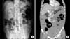
Physical examination showed moderate periumbilical tenderness without muscle rigidity. His vital signs were in normal range with blood pressure of 120/60 mmHg, heart rate of 60/min, respiratory rate of 20/min, and body temperature of 36.5℃. His bowel sound was decreased and the abdomen was soft and not distended. Laboratory test showed normal range including serum hemoglobin, amylase/lipase, liver enzyme and blood urea nitrogen (BUN)/Creatinine except for a white blood cell (WBC) count of 20,500/µL and potassium of 5.6 mmol/L. Fig. 1 shows the initial simple abdominal X-ray and abdominal CT scan. Abdominal erect X-ray views show distended small bowel loops with several air-fluid levels (Fig. 1A). We assessed that the patient's small bowel was obstructed by some cause like adhesion and not strangulated based on nonsevere abdominal pain, no fever, no tachycardia and absence of abdominal rebound tenderness. Because he had received CT scan with intravenous contrast a few hours earlier at another hospital, we delayed the CT scan with intravenous contrast. We treated him in conservative manners such as nothing per os, nasogastric tube drainage, hydration and frequent check-up of vital signs.
After 9 hours, the patient's abdominal pain worsened and we found that his abdomen was becoming distended. Blood tests showed WBC count of 16,600/µL, hyperkalemia of 7.1 mmol/L, BUN of 30 mg/dL, and Creatinine of 2.3 mg/dL. CT scan of the abdomen and pelvis was performed with intravenous contrast. CT showed mesenteric swirl signs and intestinal strangulation signs (Fig. 1B). Therefore, we performed an emergent laparotomy.
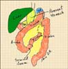
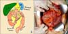
During the first operation, we found that almost all the small bowel was herniated through the Peterson's defect. Except for the proximal 20 cm of jejunum and the distal 10 cm of ileum the whole herniated small bowel and the antecolic Roux-en-Y limb were already necrotized (Fig. 2). After resection of the irreversibly strangulated small bowel, we performed an end-to-end anastomosis of the proximal jejunum and the distal ileum. The ileocecal valve was saved. And we found that there was no viable small bowel that could be connected to the remnant stomach to the remnant jejunum. We decided to link the remnant stomach with the remnant jejunum by making a new Roux-en-Y anastomosis using transverse colon segment as a new Roux-limb. At first, we made an anastomosis between the remnant stomach and the proximal transverse colon. After that, we made a distal transverse colostomy because the colon was not prepared and we wanted to prevent the risk of regurgitation of stool contents from colon into stomach and subsequent development of aspiration pneumonia in the unconscious state of the patient in the intensive care unit after the operation (Fig. 3).
We performed a second operation 32 days after the first operation. At first, we took down the transverse colostomy and anastomosed its distal end to side of the 20-cm length jejunum. Then, we cut the transverse colon at the very proximal point of gastro-colic anastomosis site and closed it. Finally, we made an end-to-end anastomosis of the ascending colon and distal transverse colon (Fig. 4).
The patient started to eat liquid diet 8 days after the second operation. At the early period of oral intake, there were some difficulties like delayed gastric emptying, constipation or frequent diarrhea. With insufficient oral intake, we supplied him additional calories with a parenteral nutritional fluid of 1,186 kcal every day. Two months after the second operation, we started the cyclic infusion of parenteral nutritional fluid so as to have the patient free from intravenous infusion at night. During that time, we supplied him with about 1,000 kcal parenteral nutritional fluid in day time via a chemoport. The amount of oral intake has increased slowly and 9 months after the second operation, he can eat a half cup of solid blended diet 3 times a day and small amount of snacks once a day. He defecates once or twice a day with the aid of a lactulose supplement, usually as loose stool or diarrhea. The patient maintained his body weight ranging from 46 to 49 kg and body mass index around 18 kg/m2 until he died. Trace elements like iron, zinc, selenium, magnesium, and vitamins were regularly checked up and we replaced the deficit. Sixteen months after his initial operation, the patient died due to bone marrow suppression and unknown cause sepsis.
DISCUSSION
Cesar Roux had performed the first human Y loop anastomosis operation for pyloric obstruction patients in 1892. But, Cesar Roux abandoned the procedure to treat benign antropyloric obstructions in 1911. After it fell into disfavor for 40 years, the Roux-en-Y reconstruction method regained popularity in many gastrointestinal surgical fields [5]. Nowadays, Roux-en-Y anastomosis operation has been performed on various conditions including hepatobiliary surgery, stomach cancer and gastric bypass surgery for morbid obesity. Roux-en-Y gastric bypass surgery is now the most common bariatric treatment for morbid obesity [4]. As the number of Roux-en-Y anastomosis operations increase annually, so too will the incidence of the complications. Known complications of Roux-en-Y anastomosis include anastomotic leak or stricture, anastomotic ulcers, small bowel obstruction, and internal hernia [4]. Especially, internal hernia is an important cause of abdominal pain after gastric bypass with an incidence ranging from 1% to 9% [3]. And it accounts for more than half the cases of small bowel obstruction after liver transplantation and after Roux-en-Y gastric bypass surgery [2] and its overall mortality is estimated at 1-2% [4]. It has been reported that laparosopic Roux-en-Y anastomosis operation showed more frequent internal herniation than open operation. With the increase of laparoscopic Roux-en-Y gastric bypass, increased incidence of internal hernia has been reported [6,7]. Internal herniation of small bowel loops after Roux-en-Y anastomosis can occur through the 3 defects, the transmesocolon defect, the mesenteric defect created by jejunojejunostomy, or the Peterson's defect (the so-called Peterson's hernia). The Peterson's defect is a potential space between a mesentery of an ascending Roux-limb and a caudal side of a mesocolon. Because the majority of internal hernias (69%) occur at the transverse mesenteric defect, the incidence of internal hernia after antecolic Roux-en-Y anastomosis is lower than retrocolic Roux-en-Y anastomosis [3,6]. In this case the patient previously underwent a near total gastrectomy and antecolic Roux-en-Y gastrojejunostomy and the mesenteric defect by jejunojejunotomy was closed with an interrupted nonabsorbable suture. The Peterson's defect was the only potential space for internal hernia.
There has been much debate about the efficacy of prophylactic closure of internal hernia defects [3,6]. Iannelli et al. [6] reviewed that routine closure with a running nonabsorbable suture resulted in reduced incidence of internal hernia in several studies. However, Greenstein and O'Rourke [3] reviewed a series with no closure and others with routine closures reporting similar incidences ranging from 0.2% to 9%, but they also recommended routine closure based on low morbidity.
In this case, the preoperative CT scan showed a swirl sign, which indicates small bowel mesenteric twisting and didn't provide any clues involving an internal hernia. However, during emergent laparotomy we found that the patient's small bowel mesentery was twisted after herniation. And, we found several reports that transmesenteric hernia following Roux-en-Y anastomosis often accompanies twisting of the small bowel mesentery and is prone to strangulation [2,4,7]. Lockhart et al. [7] reported the median amount of swirl in patients with internal hernia after Roux-en-Y gastric bypass was 180° to 270°. Mesenteric swirl sign at the abdominal CT was reported as the most predictive sign of internal hernia [4,7]. Sensitivity and specificity of mesenteric swirl in the preoperative diagnosis of internal hernia in patients who underwent Roux-en-Y gastric bypass surgery were reported to be 78-100% and 80-90%, respectively [4].
The most common clinical symptoms of a transmesenteric hernia are abdominal pain, nausea, and abdominal distension. In this case the patient didn't complain of nausea and abdominal distension was not prominent at first. Also, the patient showed moderate periumbilical pain and leukocytosis of more than 20,000/mcL but didn't show fever, tachycardia or peritoneal signs. There has been much debate about predicting strangulation with the so-called classical features like continuous abdominal pain, fever, tachycardia, peritoneal signs, and leukocytosis. Hayanga et al. [8] reviewed that it has been found consistently in both retrospective and prospective studies that these signs are not sensitive, specific, or accurately predictive of strangulation. Additionally, no blood test has ever shown practical significance in diagnosing strangulation. Furthermore, no combination of these signs can accurately predict vascular compromise. Even CT scan can't discriminate reversible strangulating obstruction from simple obstruction. CT examination is useful only for detecting the late stages of irreversible ischemia. Frazee et al. [9] reported 79% of patients with a WBC count of more than 20,000/mcL had gangrenous bowel. Roggo and Ottinger [10] reported a positive association between leukocytosis greater than 18,000/mcL and gangrenous small bowel.
Thus, it is important to remember that we cannot diagnose or exclude bowel strangulation reliably preoperatively but at least when there is leukocytosis of more than 20,000/mcL we should suspect strangulation strongly.
When herniated small bowel loops and the Roux-limb are irreversibly strangulated as in this case, a surgeon should remove the infarcted bowel and decide upon how to reconstruct the continuity of intestines. There are several reports of small bowel strangulation in patients with internal hernia who underwent Roux-en-Y anastomosis. Most of them have described simple end-to-end anastomosis of small bowel or have not described the method of reconstruction [2,6]. In this case, we rebuilt intestinal continuity from mouth to anus by making a new Roux-en-Y anastomosis utilizing a transverse colon segment as a new Roux-en-Y limb through two-stage operations. We hope this method can be the choice for surgeons who might encounter a patient with small bowel strangulation who had undergone Roux-en-Y anastomosis operation previously.

 XML Download