

 ePub
ePub Citation
 Print
Print


INTRODUCTION
Torsion of the gallbladder is a relatively rare entity that is difficult diagnose preoperatively. The condition occurs more commonly in thin elderly women [1-4], but the exact etiology is poorly understood. Although most cases are diagnosed at the time of surgery, a delay in diagnosis and treatment may result in a serious outcome. This report describes a rare case of a 36-year-old pregnant woman who presented with torsion of the gallbladder and was successfully treated with laparoscopic cholecystectomy.
CASE REPORT
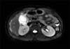
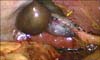
A 36-year-old pregnant woman at 17 weeks of gestation presented to the Emergency Department with a one-day history of severe epigastric pain. The abdominal pain had started as epigastric discomfort that increased in intensity and then became more localized to the right upper quadrant. The pain was sharp and constant in nature. Physical examination revealed a thin woman (height, 165 cm; weight, 56 kg) with a blood pressure of 123/64 mmHg, a pulse of 80 beats/min, and a body temperature of 36.6℃. The abdomen was tender in the right upper quadrant. No guarding and rebound tenderness were noted. The laboratory data showed a white blood cell count of 9,700/mm3, a hemoglobin level of 11.8 g/dL, and the platelet count was 152,000/mm3. Other blood chemistry parameters including liver function test were unremarkable. Abdominal US demonstrated a distended gallbladder without stones but with mild wall thickening. Magnetic resonance imaging (MRI) demonstrated a slightly distended gallbladder with a markedly edematous and multilayered wall (Fig. 1). A diagnosis of acute cholecystitis was made, and antibiotics were administered; however, the patient's symptoms worsened. On the third hospital day, laparoscopic cholecystectomy using three-port was performed under the impression of an acalculous cholecystitis. The gallbladder was gangrenous and the body of the gallbladder had rotated 180° clockwise on the gallbladder mesentery (Fig. 2). The gallbladder was supported by the mesentery which was attached only to the proximal body of the gallbladder, thereby allowing the gallbladder to hang free. The gallbladder was untwisted and then removed without difficulty. Histopathological examination showed an acute gangrenous inflammation with extensive infarction. The postoperative course was uneventful and the patient was discharged on the 4th day after surgery.
DISCUSSION
Torsion of the gallbladder is a condition that is difficult to diagnose preoperatively. The condition can occur at any age and in either sex, but has a predilection for the elderly, with a 3:1 female-to-male ratio. The increased incidence of this condition may be attributable to increasing life expectancy.
The etiology of torsion of the gallbladder is uncertain. However, several hypotheses have been postulated as the mechanism. There are two requirements for torsion of the gallbladder: an anatomic configuration, allowing rotational gallbladder mobility, and a triggering event that results in the gallbladder twisting around the cystic duct as an axis point [5]. The anatomic configurations necessary for torsion of the gallbladder are well documented, but the triggering event is poorly understood. Two types of anatomical variants have been implicated in the majority of cases, as well as a third, less common condition [5-7]. The first may be related to the congenital deformity. Between the 4th and 7th weeks of embryological development, the pars cystica forms from the hepatic diverticulum. Abnormal migration, with an absence of a gallbladder mesentery, creates a "free-floating gallbladder" or "mobile gallbladder". The second, long mesentery type, as in our case, occurs by generalized visceroptosis. The mesentery of the gallbladder and cystic duct relax and elongate with advancing age, althouth our case was relatively young, creating a mobile situation. Atrophy of the liver, loss of visceral fat and elasticity with aging, weight loss and spinal deformities may place thegallbladder in a more dependent position, with a predisposition to torsion. An extremely rare third variant consists of a normally fixed gallbladder to a mobile liver lobe free of its coronary and triangular ligaments.
Carter et al. [8] described two types of torsion, incomplete torsion, with a rotation of less than 180 degrees and with gradual onset, and complete torsion, as in our case, with a rotation more than 180 degrees with acute onset. Complete torsion occludes both the bile and blood flows, whereas incomplete torsion occludes only the bile flow. Complete torsion interferes with the blood supply to the organ, and if this is unrelieved, gangrene will develop.
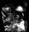
Nonspecific symptoms, nonspecific findings on physical examination and its rarity make the preoperative diagnosis difficult. Despite the recent advances of the radiologic imaging modalities, making the correct preoperative diagnosis of gallbladder torsion is challenging and most cases are diagnosed at the time of surgery. Ultrasonography (US) and computed tomography are useful modalities and some findings can help clinicians diagnose gallbladder torsion preoperatively when they have a suspicion of this disease. The decisive findings were the left-sided enlarged gallbladder, thickened gallbladder wall with no enhancement effect, and a cystic duct located on the right side of the gallbladder [9]. On US examination, another specific sign observed with torsion of the gallbladder is the presence of the gallbladder outside its normal anatomic fossa, and inferior to the liver with an echogenic conical structure [9]. Usui et al. [10] reported the magnetic resonance cholangio-pancreatography findings of gallbladder torsion to be as follows: 1) a v-shaped distortion of the extrahepatic bile ducts due to traction by the cystic duct, 2) tapering and twisting interruption of the cystic duct, 3) a distended and enlarged gallbladder that deviates to the midline of the abdomen, and 4) a difference in intensity between the gallbladder and the extrahepatic bile ducts and the cystic duct. Unfortunately, although the preoperative diagnosis of gallbladder torsion was missed in our case, retrospecitve review of a preoperative MRI scan provided some diagnostic clues of a markedly edematous wall of gallbladder, sightly v-shaped distorsion of (Fig. 3) and no visualization of gallbladder in magnetic resonance cholangiography images. The absence of gall stones in a gallbladder with signs of cholecystitis suggests a torsion of the gallbladder since an acalculous cholecystitis is very rare in otherwise healthy patients.
Emergency cholecystectomy should be performed when gallbladder torsion is suspected. A laparoscopic approach is now recommended as the first choice of treatment due to the increased experience with laparoscopic cholecystectomy. An early diagnosis and performing prompt cholecystectomy for this disease is important in order to avoid the complications of gangrene and perforation, and to reduce the resultant mortality.
In conclusion, torsion of the gallbladder is a rare clinical entity that requires immediate surgical treatment. Therefore, it is important to keep in mind the gallbladder torsion in the differential diagnosis from acute cholecystitis when the patient has an acute onset of abdominal pain with minimal episodes of fever or jaundice and a severely distended gallbladder. Prompt cholecystectomy via a laparoscopic approach should be performed.
 XML Download