

 ePub
ePub Citation
 Print
Print


INTRODUCTION
Thyroid stimulating hormone (TSH) level represents thyroid function sensitively and is used for evaluation of thyroid nodules. TSH is also well known as a thyrocyte growth factor. Recently, several investigators studied correlations between preoperative serum TSH levels and thyroid carcinoma status. Some of them showed significant correlation but others did not.
Serum TSH level can be affected by various confounding factors such as the age of the patients, medication history, diseases affecting thyroid function, pregnancy, obesity, etc. We believe that these inconsistent results came from different considerations of these confounding factors.
In this study we therefore evaluate more direct correlations between the preoperative serum TSH level and thyroid nodule status by excluding these confounding factors.
METHODS
From January 2009 to December 2010, 383 patients underwent thyroid surgery for nonfunctioning thyroid nodule at Chungbuk National University Hospital. Two hundred fifty-seven patients who had confounding factor(s) affecting serum TSH level were excluded: patients who had auto-antibodies (such as antithyroglobulin-Ab, antithyroid peroxidase [microsomal]-Ab, or anti-TSH receptor-Ab), abnormal serum TSH levels, histologically proven Hashimoto thyroiditis, and a history of taking synthyroid or estrogen, patients who showed a marked heterogenous echogenecity on ultrasonography and patients whose medical records did not contain information about these factors. The remaining 126 patients were included in this retrospective analysis.
Average age of patients was 45.4 ± 10.6 years and the male to female ratio was 1:2.9. Eleven patients had benign nodules only including nodular hyperplasia and benign thyroid tumors. One hundred fifteen patients were diagnosed with papillary thyroid carcinoma (PTC). The maximal carcinoma diameters were not more than 5 mm in 34 patients, more than 5 mm but not more than 10 mm in 66 patients, more than 10 mm in 15 patients. SPSS ver. 16.0 (SPSS Inc., Chicago, IL, USA) used for a statistical analysis with significant results defined as P < 0.05.
RESULTS
Demographic characteristics of patients
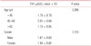
There were no significant differences in mean serum TSH concentrations according to age groups. Mean serum TSH concentration was 1.79 ± 0.79 µIU/L under age of 45 years, 2.01 ± 0.84 µIU/L between age of 45 years and 60 years, 1.78 ± 0.95 µIU/L over age of 60 years. In terms of gender, there was no significant difference either: 1.67 ± 0.83 µIU/L for male and 1.94 ± 0.82 µIU/L for female (Table 1).
Nodular characteristics
There were no statistically significant differences in mean serum TSH concentrations between patients who had benign nodule(s) (1.55 ± 0.65 µIU/L) and malignant nodule(s) (1.87 ± 0.83 µIU/L) (P = 0.22). Maximal tumor diameter, volume of the biggest nodule or total volume of all nodules did not correlate with preoperative serum TSH concentrations (P = 0.414).
Patients who had PTC
PTC was diagnosed in 115 patients. Neither the size(s) nor volume(s) of malignant tumor(s) showed any meaningful correlation with preoperative serum TSH concentration. However, preoperative serum TSH levels and advanced stages of PTCs might correlate closely as some previous investigators suggested. We therefore evaluate its correlation with TNM stage (American Joint Committee on Cancer 7th, 2010). We presumed M0 when there was no clinical or imaging evidence of distant metastasis and undetectable serum Tg levels during TSH suppression and stimulation in the absence of interfering antibodies excluding locoregional recurrences at the final follow-up. There was no M1 patient.
T stage
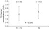
Fifty-five, three, and fifty-seven patients were in T1, T2, and T3 clinical stage, respectively. As the number of patients in T2 stage was too small, T1 and T2 stage were combined as a group for statistical analysis. Mean serum TSH concentrations were 1.72 ± 0.79 µIU/L in T1 + T2 group and 2.02 ± 0.85 µIU/L in T3 group, respectively. The difference was statistically significant (P = 0.046) (Fig. 1).
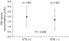
T3 group is heterogenous because both the size of primary tumors (over 4 cm in diameter) and presence of minimal extrathyroidal extension are considered. As the size of primary tumors was evaluated early, correlation between serum TSH levels and presence of extrathyroidal extension was investigated. Mean serum TSH concentration of patients who had tumors with extrathyroidal extension was 2.02 ± 0.84 µIU/L, which was higher than that of patients who did not, 1.72 ± 0.79 µIU/L. However, the difference was not statistically significant (P = 0.059) (Fig. 2).
N clinical stage
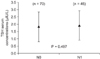
Seventy, forty-two, and three patients were in N0, N1a, and N1b stage, respectively. Mean preoperative serum TSH concentration was 1.81 ± 0.77 µIU/L in N0 group, 1.86 ± 0.86 µIU/L in N1a group, and 3.26 ± 0.63 µIU/L in N1b group, respectively. Although the number is small, patients in N1b group showed significantly higher serum levels than the others (P = 0.012). When we compared N0 group with N1 (N1a + N1b) group, there was no significant difference (P = 0.497) (Fig. 3).
TNM stage
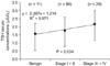
Eighty-three, three, twenty-seven, and two patients represented stage I, II, III, and IV, respectively. Mean preoperative serum TSH concentration was 1.76 ± 0.82 µIU/L in stage I, 1.92 ± 0.68 µIU/L in stage II, 2.06 ± 0.75 µIU/L in stage III, and 3.60 ± 0.28 µIU/L in stage IV, respectively. Mean preoperative serum TSH concentration in patients who had benign nodule(s) was 1.55 ± 0.65 µIU/L. The more advanced the disease status, which was from benign to stage IV PTC, the higher mean preoperative serum TSH concentration was (P = 0.008). The trend was maintained when we compared serum TSH levels in 3 groups, that is, benign group, early stage of PTC (stage I and II), and late stage of PTC (stage III and IV). Mean preoperative serum TSH concentration was 1.55 ± 0.65 µIU/L, 1.77 ± 0.81 µIU/L, 2.16 ± 0.82 µIU/L, respectively (P = 0.034) (Fig. 4).
DISCUSSION
Measurement of serum TSH concentration is the initial evaluation of a patient with a thyroid nodule. TSH is well known for a growth factor that acts on thyroid tissue. There are many studies about its relation to thyroid tumors. There is, however, a variety of opinions concerning how TSH affects thyroid tumors.
Many studies have reported a weak association between serum TSH concentration and thyroid cancer, whereas Matsuo et al. [1] reported that mutational activation of the intracytoplasmatic domains of the TSH receptor is not a significant mechanism of thyroid tumorigenesis.
Derwahl et al. [2] and Mazzaferri [3] reported its relation to other definite growth factors such as insulin-like growth factor-I in vitro. Shi et al. [4] suggested that decreased TSH receptor gene expression levels are associated with thyroid cell dedifferentiation. Administration of iodine to patients who have goiter with high serum TSH concentration in iodine deficiency areas can induce decreasing goiter volume by lowering of serum TSH level [2]. Many investigators have shown that the administration of thyroid hormone can prevent the growth of pre-existing thyroid nodules and the formation of new nodules [2,5-7]. It is also supported by the fact that patients who have autoimmune thyroid disorders such as Hashimoto thyroiditis showed an increased risk of thyroid cancer by 2.77 times [8]. THS suppressive therapy is a well-known adjuvant therapy to prevent recurrences in follicular cell derived differentiated thyroid cancer (DTC) patients [9], although further studies on specific mechanisms are still required.
In this context, correlations between preoperative serum TSH level and DTC have recently been investigated. Polyzos et al. [10] and Fiore et al. [11] found that the risk of PTC was lower in patients who had serum TSH concentrations below the normal reference range than patients who did not. In a study of 1,500 patients who underwent fine needle aspiration biopsy, Boelaert et al. [12] reported that the risk of malignancy increases when patients have single thyroid nodule with serum TSH concentrations over 0.9 mIU/L. Haymart et al. [13] supported this finding. He reported that the likelihood of malignancy of thyroid nodule was 16%, 25%, 35%, and 52% when serum TSH concentration was below 0.06 mIU/L, 0.40 to 1.39 mIU/L, 1.40 to 4.99 mIU/L, and 5.0 mIU/L or greater, respectively. He also reported that higher serum TSH level in thyroid nodule patients was associated with advanced tumor stage and poor prognosis. On the contrary, Gerschpacher et al. [14] recently reported that there was no significant difference in mean preoperative serum TSH level between patients with PTCs and the control group.
We believe that these inconsistent results came from different considerations of confounding factors which affect serum TSH levels such as the age of the patients, medication history, diseases affecting thyroid function, pregnancy, obesity, etc [15].
In this study, we therefore tried to evaluate more direct correlations between the preoperative serum TSH level and thyroid nodule status by excluding these confounding factors. After excluding as many confounding factors as possible, we could find a meaningful relation of preoperative serum TSH concentration to the clinical stage of PTC but failed to show its relation to the size of nodule whether it is benign or malignant. Mean preoperative serum TSH concentrations are significantly higher in T3 group than in T1 + T2 group (2.02 ± 0.85 µIU/L vs. 1.72 ± 0.79 µIU/L, P = 0.046). As T3 group is heterogenous, subgroup analysis according to extrathyroidal extension was done. Mean serum TSH concentration of patients who had tumors with extrathyroidal extension tends to be higher than that of patients who did not (2.02 ± 0.84 µIU/L vs. 1.72 ± 0.79 µIU/L, P = 0.059). Although the number is small, patients in N1b group showed significantly higher serum levels than N0 or N1a group (3.26 ± 0.63 µIU/L vs. 1.81 ± 0.77 µIU/L vs. 1.86 ± 0.86 µIU/L, P = 0.012). The more advanced the disease status was from benign to stage IV PTC, the higher mean preoperative serum TSH concentration was: 1.55 ± 0.65 µIU/L in benign nodule(s), 1.76 ± 0.82 µIU/L in stage I, 1.92 ± 0.68 µIU/L in stage II, 2.06 ± 0.75 µIU/L in stage III, and 3.60 ± 0.28 µIU/L in stage IV (P = 0.008).
There are limitations in this study. The study population was insufficient and all factors that affect TSH were not completely excluded, so more studies are required to achieve more solid conclusions. Despite these limitations, we might suggest that if the preoperative TSH serum concentration is high while the other factors affecting serum TSH concentration are excluded, the possibility of advanced PTC should be considered regardless of the size of nodules.
In conclusion, even in the normal range of serum TSH concentrations, higher levels of preoperative TSH serum concentrations might be related to advanced TNM stages of PTC if there are no factors affecting TSH serum concentration, no ultrasonographic findings suggestive of thyroiditis, and no thyroid auto-antibodies.
 XML Download