

 ePub
ePub Citation
 Print
Print


INTRODUCTION
Nevus lipomatosus cutaneous superficialis (NLCS) is a relatively rare benign abnormality of the connective tissue that is characterized by the presence of mature ectopic adiopocytes in the dermis classified into two clinical subtypes: multiple and solitary [1,2]. Mehregan et al. [3] proposed the term 'pedunculated lipofibroma' for the solitary form of NLCS.
We report here on a case of a heart-shaped pedunculated lipofibroma occurring on the coccygeal area in the neonatal period.
CASE REPORT
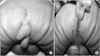
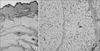
A 3-day-old male neonate was referred to Department of Pediatric Surgery, Seoul National University Children's Hospital with a mass on the coccygeal area. The neonate was delivered via normal spontaneous vaginal delivery with 37 weeks gestation, as the second of twins, and measured a weight of 1,810 gm with normal Apgar scores. Physical examination revealed a 1.5 × 1.8 cm sized soft, flesh-colored, heart-shaped mass with a smooth surface. The mass showed a pedunculated shape and was connected to the skin and subcutaneous tissue of coccygeal area by a stalk (Fig. 1A, B). Ultrasonography and magnetic resonance imaging showed the hyper-echoic soft tissue mass was not communicating with the spinal cord. The mass was excised with ligation of the feeding vessel, which was surrounded with fibrous tissue at the core of the stalk. The pathologic exam was compatible with pedunculated lipofibroma. There were many collections of mature adipose cells trapped between bundles of collagen in the dermis with no evidence of accompanying melanocytes (Fig. 2A, B)
DISCUSSION
In 1921, Hoffmann and Zurhelle [4] first reported a case of NLCS. NLCS is a rare benign hamartomatous condition that is characterized by ectopic mature adipose tissue in the dermis [4,5].
The pathogenesis of peduculated lipofibroma is unknown despite several theories. The proposed theories are degenerative changes in the collagen and elastic tissue, displacement of subcutaneous adipose tissue into the dermis, and origination and differentiation from the walls of dermal vessels [2,6].
There are two clinical subtypes of NLCS: a multiple form (the classic type) and solitary form [2]. The classic form presents as grouped soft, smooth papules or nodules at birth and develops during the first three decades of life. They often coalesce to form plaques. Their surfaces are usually smooth but may be verrucous or cerebriform. These are most commonly seen in the pelvic girdle, with a predilection for the gluteal region. On the other hand, the solitary form usually presents as a single nodule or papule in adults and can appear at any site, including the lower trunk, knee, axillae, arm, ear or scalp [7].
The diagnosis of peduculated lipofibroma can be confused with other benign papillomas due to their characteristic appearance. The diagnosis of peduculated lipofibroma can be confirmed by the histopathologic findings showing isolated groups of ectopic mature adipocytes within the reticular dermis [1,8].
Treatment is generally not necessary except for cosmetic reasons. Surgical excision may be applied if there are cosmetic and functional problems, and recurrence has not been reported.
We observed a rare case of neonatal pedunculated lipofibroma with a prominent stalk; such a lesion usually develops in adults. We treated the mass with total excision.
 XML Download