

 ePub
ePub Citation
 Print
Print


INTRODUCTION
Advances in surgical intensive care have enabled patients with severe abdominal pathologies to survive. En bloc tumor resections, previous surgeries, intraabdominal catastrophes, necrotizing fascial infections, or traumatic abdominal injuries cause abdominal wall defects in these kind of patients [1,2]. The aims of reconstruction in such defects are full restoration of abdominal wall function, including muscular support, prevention of visceral eventration, and adequate soft tissue coverage [3].
Wide undermining of the skin and subcutaneous tissue usually permits primary wound closure. In a minority of patients, lateral migration of the rectus abdominis muscles from contraction of the flank muscles, and concomitant visceral sac protrusion, result in loss of abdominal domain that make this approach impossible [2]. Generally, two methods are used for the treatment of abdominal wall defects that are not amenable to tensionless approximation of the natural tissues. The first method is to bridge the defect with the patients' own tissue, synthetic products, or a composite material; and the second one is to reapproximate the natural tissue after utilizing relaxing incisions or preoperative measures like tissue expansion or progressive pneumoperitoneum [3].
Synthetic meshes, autologous tissue flaps, mostly myocutaneous flaps such as tensor fasciae latae, rectus femoris, rectus abdominis, and latissimus dorsi, with or without incorporation of synthetic meshes, pedicled omentum flaps, some biological materials like human acellular dermal matrix, were introduced in hernia repair and abdominal wall reconstruction [4-6]. Abdominal wall reconstruction with prosthetic mesh can be safe and effective if well-vascularized soft-tissue coverage is provided.
Tissue expanders have been used in this clinical situation to provide soft-tissue coverage and to restore abdominal domain by increasing both the size and the vascularity of the donor tissue. Tissue expansion produces a strong, vascularized capsule around the expanders. Our patient's vascularized capsule, combined with the existing rectus sheath, was used to reconstruct abdominal wall defects [7]. Tissue expanders placed in the subcutaneous space, abdominal wall intramuscular spaces (between the internal oblique and transverse abdominis muscles), intermuscular sites (between the external and internal oblique muscles), and finally intraabdominally, have been reported as a method to increase the size of potential skin or muscle flaps before closure and also for stable coverage of prosthetic material [3,8].
Simplicity, safety and efficacy should be the main considerations in the management. In this report we describe our experience in treating a patient with large abdominal wall defect by staged abdominal wall reconstruction utilizing prosthetic mesh in conjunction with tissue expanders.
CASE REPORT
A 41-year-old male patient presented to our emergency department with abdominal pain. Exploratory laparotomy performed through a subumbilical incision showed perforated appendicitis with intraabdominal abscess of 1,500 mL. In addition, there was a large hemangioma occupying 80% of the liver. After surgery he developed intraperitoneal sepsis and in order to prevent abdominal compartment syndrome, he was reoperated and left with "open abdomen". Several open abdomen lavages were performed and his abdominal wall defect was allowed to granulate.
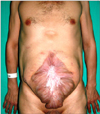
After epithelization of the defect approximately ten months later (Fig. 1), reconstruction of the abdominal wall was planned. Skin incisions were made 2 cm lateral to the defective area. Two rectangular tissue expanders (LS 82 2000, Laboratoires Sebbin, Boissy-l'Aillerie, France) of 2,000 mL volume (230 × 130 × 70 mm) were placed in the subcutaneous pockets created just above the anterior rectus sheath and adjacent to the abdominal wound bilaterally to reconstruct the defect of 23 × 37 cm in size. The ports of the tissue expanders were placed on the thoracic cage region.
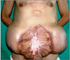
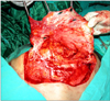
Each of the tissue expanders were inflated with 200 mL of saline at the time of the initial operation. Over the next several weeks, the expanders were gradually inflated to a total volume of 2,350 mL each over a period of four months (Fig. 2). After four months, the patient returned to the operating room for definitive repair of the defect. The expanders were removed (Fig. 3) and since the fascial defect was too large to close primarily, utilizing prosthetic mesh (expanded polytetrafluoroethylene; GORE-TEX soft tissue patch) (Fig. 4) in conjunction with tissue expanders, the abdominal wall defect was reconstructed. Expanded skin flaps were advanced toward the midline and closed in layers without tension. A more acceptable midline scar was then obtained. Suction drains were placed bilaterally and removed three days later.
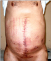
The postoperative period of the patient was uneventful and he was discharged on the 4th postoperative day. During follow-up for ten months, he showed no signs of mesh infection or extrusion, and did not develop recurrent hernia, ulceration or enteric fistula. The tissue expansion process was well tolerated by the patient (Fig. 5).
DISCUSSION
Treatment of large ventral hernias is difficult, partly due to a lack of available abdominal wall, and owing to the difficulty in finding sufficient space for the abdominal contents after reposition. The abdominal wall is a multilayered structure composed of several different tissues. The myofascial layer is the major supportive part of the abdominal wall, which is important for abdominal contents protection and dynamic function [4].
There is no consensus about which technique or material is suitable for a certain kind of defect. Selection should be made mainly on the basis of the size, location and depth of the defect, wound bed preparedness and the medical status of the patient [1,4]. When assessing the patient initially, a complete history, physical and laboratory workup are essential with the involvement of multiple specialties, including general surgery, internal medicine, nutrition, pulmonology, and infectious disease [1]. Although multiple strategies are advocated, even for different situations, it requires careful planning to determine the optimal choice in each case [2].
The goals of abdominal reconstruction are; restoration of function and integrity of the musculofascial abdominal wall, prevention of visceral eventration, and provision of dynamic muscle support [1]. There are three types of abdominal wall defects. Type I involves only the loss of skin and can be corrected with primary suture; type II is a myofascial defect with intact skin coverage for reconstruction of which a myofascial substitute is needed; and type III is a myofascial defect without skin coverage that needs autologous tissues and synthetic or biological mesh [4]. Our patient is included in type III and was repaired by prosthetic mesh and tissue expanders.
Ideally, the abdominal wall is reconstructed by mobilizing local tissue and using existing fascia. However, primary closure performed under tension results in tissue ischemia, wound dehiscence, herniation, pulmonary complications secondary to increased intraabdominal pressure. Reduced diaphragmatic excursion is associated with high morbidity and mortality rates. The use of prosthetic mesh in conjunction with tissue-expanded skin provides a durable abdominal closure and is technically simpler than flap closure methods.
With minimal donor site morbidity relative to most musculofascial techniques, tissue expansion provides well-vascularized skin and soft tissue over the prosthetic mesh, allows excision of unsightly scars and skin grafts and obtains excellent color and texture match [2]. Subcutaneously placed tissue expanders have been successfully used in surgical situations with the primary intent of expanding skin to achieve wound closure, including adult patients who had loss of the abdominal wall from either traumatic or infectious events and in pediatric patients with abdominal wall defects [8]. Subcutaneous placement under healthy skin provides sufficient skin and subcutis to be approximated and allows uncomplicated placement of expanders and ample recruitment of soft-tissue coverage, with prosthetic mesh being used to close fascial defects [3]. But, the relatively superficial location of the expander can cause thinning of the skin and result in extrusion that necessitates early removal and placement of a new expander at a later date [8]. Subcutaneous tissue expansion does not result in any alteration to the structure of the Musculofascia labdominal wall but can serve as an adjunct to bridge these defects [3].
Subfascial placement of expanders permits expansion of muscle and fascial layers to provide full-thickness autologous abdominal wall reconstruction, but at the cost of slightly more difficult dissection and subsequent removal. Placing expanders in the plane between the transverse and internal oblique muscles is risky because this area contains the nervous and arterial supplies for these two muscles and the rectus [3]. As a prevention, the vertical incision made in the posterior layer of the internal oblique aponeurosis should be minimal in length and performed with caution [8]. From an anatomic point of view, the plane between the external oblique and internal oblique muscles seems to be the most convenient to use, but, it is a time-consuming, expensive form of therapy, as the process of gradual expansion takes several weeks resulting in limited enlargement of the structures needed [3].
De Ugarte et al. [8] presented a novel approach to using tissue expanders placed within the intramuscular layers of the abdominal wall of a patient with a giant omphalocele, allowing creation of an outer layer composed of skin and internal and external oblique muscles. Intraabdominal placement of tissue expanders offers the advantage of creating "tissue flaps" that include all layers of the abdominal wall, including skin, muscle, and peritoneum. On the other hand, expansion of the abdominal cavity can increase intraabdominal pressure, impair respiratory function, cause visceral ischemia and increase the risk of displacement of viscera [8].
The early use of tissue expanders to increase the amount of tissue available for coverage would be limited by the presence of intestinal contamination, stomas, and drains [9]. Before starting filling of the expander, a period of wound healing is usually awaited for three weeks to prevent expander exclusion. Tissue expanders can become infected or exposed during the course of expansion with a related complication rate of less than 15% . The predicted surface area gain after completion of tissue expansion often falls short of the clinical requirements [10]. Due to the reversible phenomena of expanded skin to retract after removal of the expanders, overexpansion (10% to 20% over implant capacity) is necessary. Instead of resorting to serial expansions, overexpansion techniques help in reaching the desired dimensions and avoiding a shortage of donor site for expansion, and eventually reconstruction can be completed in one-stage [10]. It appears safe without risk of implant failure at least to 5 to 10 times the vendors' stated maximum volume.
As a conclusion, the ideal reconstruction of abdominal wall should provide tension-free repair and dynamic muscle support if possible. Tissue expanders can be used as an additional therapeutic modality in the treatment of complex abdominal wall defects not amenable to delayed primary closure. The location for placement of a tissue expander depends on the medical condition and specific reconstructive needs of the patient. This method has provided reliable and durable abdominal wall closure with adequate soft-tissue coverage of prosthetic mesh as well as partially restoring abdominal domain. It also minimizes the potential mesh related complications, and is aesthetically superior to that achieved with skin graft and muscle flap techniques. When the size, place, and depth of the expander is chosen carefully, and the expanding process and antibiotic prophylaxis is managed properly, this staged method of abdominal wall reconstruction is safe and simple regarding the literature and our experience. Although the expansion process takes longer and, in some countries, the use of expanders has some medical and socioeconomical problems (cost, insurance coverage problems, etc) it allows maximization of patient healing and provides good functional and aesthetic results as in our patient.

 XML Download