

 ePub
ePub Citation
 Print
Print


INTRODUCTION
Fecal incontinence is a socially disabling disorder that profoundly impacts on all aspects of quality of life. It is thought to effect 2% to 15% of community-dwelling adults [1,2]. Fecal incontinence is considered to be an aging disease because its prevalence increases to 13% among people older than 50 years [3], and is an iatrogenic disease that develops in 60% of rectal cancer patients after intersphincteric resection [4]. However, the treatment of fecal incontinence is an area of uncertainty because data from randomized trials are lacking to establish optimal approaches for its treatment [5]. Although injection of a bulking agent has been considered as a minimally invasive treatment [6], its efficacy is limited by several factors, including reabsorption, migration [7], fat embolism, and granuloma formation [8].
Approaches aimed at regenerating functional muscle have been proposed as a therapeutic tool for fecal incontinence [9]. Some pioneers in the field of coloproctology have shown that autologous adipose stem cells may be used to treat fistulas in patients with Crohn's disease; these cells did not migrate or cause embolism [10]. Several reports showed that injection of cells into the anal sphincter improved functional outcomes in rat [11-14] and rabbit [15] models of fecal incontinence. Additionally, several clinical trials have tested whether the injection of myoblasts improves incontinence, but there was no histologic evidence demonstrating the induction of functional muscle [16]. We consider that cell therapy alone without a bioscaffold produced by bioengineering technique would provide limited functional and structural improvements in fecal incontinence patients with large structural deformities [17], which differs from urinary incontinence, which shows less marked structural deformities.
Until recently, no studies have used bioengineering techniques to improve fecal incontinence. In our previous study, the use of polycarprolactone porous beads [18] facilitated smooth muscle regeneration and improved electrical contractility in vitro evaluation [19], which indicates that these beads may be useful as an injectable bioactive bulking agent for treating incontinence [20]. Therefore, the present study was designed to determine whether the injection of porous polycaprolactone beads containing autologous myoblasts could improve sphincter function in a dog model of fecal incontinence.
METHODS
Animal care and experimental design
This protocol was reviewed and approved by the Animal Care and Use Committee of Seoul National University Bundang Hospital (Protocol No. 64-2010-037). Normal 10-week-old female mongrel dogs (19 to 22 kg) were used in these experiments. The animals were intubated and mechanically ventilated with a Zoletil (Virbac, Carros, France) + Rompun (Bayer Korea, Seoul, Korea) mixture to maintain surgical anesthesia. All procedures were performed in the lithotomy position. Ketoprofen was used for pain control and cefazolin was injected intravenously before and immediately after surgery. In each animal, sphincter injury was induced by partial extraction of approximately 25% of the posterior internal/external anal sphincter using electrocautery.
After inducing anal sphincter injury, the dogs were randomly allocated to two groups and received without (n = 5) or with (n = 5) an injection of porous polycaprolactone beads containing autologous myoblasts into the injured sphincter. In the autologous myoblast bead injected group, a muscle biopsy of 1 cm3 in size was taken from gastrocnemius muscle to isolate myoblasts at the time of the surgery to injure the anal sphincter. Three weeks later, the porous polycaprolactone beads containing the autologous myoblasts were injected into the site of injury in the healing process.
Compound muscle action potentials (CMAP) were measured and manometry of the anal sphincter was performed just before injury and 3 months after the intervention. The anal sphincter was removed for histopathological evaluation.
Electrophysiological assessment
We stimulated the inferior rectal branch of the pudendal nerve located 3 cm proximal to the anal verge using a stimulator constructed in-house with a needle evoked potential electrode (Oxford Instrument Medical Systems, Oxfordshire, UK). CMAPs were recorded from active and reference electrodes using a Teflon-coated monopolar needle electrode (37 mm long, 0.30 mm diameter, Oxford Instrument Medical Systems) inserted into the superficial external anal sphincter muscles. A portable two-channel electromyography/nerve conduction velocity system (Medelec Synergy, Oxford Instrument Medical Systems) was used with a filtering frequency of 10 Hz to 10 kHz, sweep speed of 1 ms/division and sensitivity of 1 mV/division. The mean amplitude and area of the CMAP were measured for five pudendal nerve conduction studies in each dog.
Anal pressure measurement
Anal sphincter pressure was measured by real-time pressure monitoring (Creta Co., Seoul, Korea) using a pressure balloon sensor (unit, mmHg; sensitivity 0.06 kPa, sensitivity 5 mV, threshold 1.5 V; Boston Scientific, Place Natick, MA, USA) introduced into the animal rectum. The sensor probe was inserted about 1.5 cm into the anal orifice to be placed in the middle of the anal sphincter muscle. The baseline resting pressure was determined as the mean pressure in the noncontractile state after balloon insertion. The squeezing pressure was defined as the maximal pressure generated by the sphincter during tetanic stimulation of the pudendal nerve (50 Hz, 50 mA stimulus intensity, 5 seconds) and was measured at least three times in each dog.
Isolation, culture and identification of myoblasts
Myoblasts were purified by the modified preplate technique, as previously described [21,22]. To characterize the cultured cells, the cells were cultured on chamber slides and fixed in cold methanol for 10 minutes. The cells were then incubated for 1 hour with rabbit polyclonal anti-Pax7 primary antibody (1:300; Lifespan Bioscience, Seattle, WA, USA), followed by labeling with polymer-horseradish peroxidase (HRP) antimouse secondary antibody (EnVision + System-HRP [3.3'-diaminobenzidine {DAB}], Dako, Glostrup, Denmark). Some cells were stained without the primary antibody as a negative control. Nuclei were stained with 3.3'-diaminobenzidine chromogen solution (Dako).
For the fusion assay, the purified cells were resuspended in differentiation medium (Dulbecco's modified eagle medium/F12 (1:1) + 2% horse serum + 1% penicillin/streptomycin). Myoblast differentiation was examined immunocytochemically using myosin heavy chain (MHC) monoclonal antibody (1:25; Novus Biologicals, Littleton, CO, USA) and α-smooth muscle actin (SMA) monoclonal antibody (prediluted, Dako). The cells were visualized under a microscope by a pathologist blinded to the study design.
Preparation of porous beads
Polycaprolactone/Pluronic F127 porous beads (size range, 200 to 300 µm) were prepared as previously described [18-20]. Briefly, polycaprolactone (Mw 43,000-50,000; Polysciences, Warrington, PA, USA) and Pluronic F127 (EG99PG65EG99, Mw 12,500; BASF, Parsippany, NJ, USA) were used to fabricate the porous polycaprolactone beads. The beads were first fabricated by isolated particle melting to generate nonporous beads, followed by melt-molding particulate-leaching to generate porous beads, as described elsewhere [18]. Ultrapure grade water (>18 mΩ) was purified using a Milli-Q purification system (Millipore Co., Billerica, MA, USA). For animal studies, the porous beads were sterilized by ethylene oxide, and the Pluronic F127 solution was autoclaved before use.
In vitro cell culture in porous beads and transplantation
As previously described [19], to seed the cells into the porous beads, the beads were immersed in a needle tip-stopped syringe filled with 2 mL of the cell suspension (cell density, 3 × 10⁷ cells/mL). The cell suspension was transferred into the porous beads (10 mg) under negative pressure when the syringe piston was pulled out. The beads were maintained for 2 hours at 37℃ in an incubator with a humidified 5% CO2 atmosphere for the cells to adhere to the beads. To ensure even delivery of the cell-seeded porous beads during injection, the beads were uniformly dispersed in Pluronic F127 solution (in phosphate buffered saline, 25 wt%; gelation temperature, about 17℃) with a bead:solution ratio of 1:12 (w/v). The cell-seeded porous beads were injected into the injured tissue using a syringe fitted with a 19 G needle. The beads in the gelled state of Pluronic F127 at room temperature could be homogeneously injected through the syringe needle without blocking the needle.
Immunohistochemistry and histopathology
At the end of each experiment, the dogs were euthanized by an intravenous overdose of KCl after intramuscular injection of Zoletil/Rompun (4.4 + 2.2 mg/kg). After the dogs were euthanized, the rectum and anal canal were dissected, immersion fixed in neutrally buffered formalin, embedded in paraffin, sectioned, and stained with hematoxylin and eosin (H&E).
After H&E slide review, representative blocks were selected. Four-micrometer sections were cut from the original blocks and stained for MHC mouse monoclonal antibody (1:200; Novus Biologicals) and SMA mouse monoclonal antibody (prediluted, Dako). The slides were deparaffinized, and hydrated in graded ethanol. Antigen retrieval was performed by incubating tissue sections in a microwave oven in 0.01 M citrate buffer (pH 6.0) for 15 minutes. The slides were incubated with primary antibody, followed by labeling with polymer-HRP antimouse secondary antibody (EnVision + System-HRP [DAB], Dako). For detection, 3.3'-diaminobenzidine chromogen solution (Dako) were applied. Normal skeletal and smooth muscle cells were used as internal positive controls, and fibroblasts and epithelial cells were used as internal negative controls. Some slides were stained without the primary antibody as a negative control.
Statistical analysis
Data are expressed as the median and interquartile range. Paired t-tests and Wilcoxon signed rank test were used for comparisons of functional outcomes measured before sphincter injury and at 3 months after injury in the dogs without the injection of porous polycaprolactone beads containing autologous myoblasts. Functional outcomes were compared by analysis of covariance (ANCOVA), with the corresponding baseline measurement as the covariate. All statistical tests were two-sided and P < 0.05 was considered significant. All analyses were performed using the IBM SPSS ver. 18.0 (IBM Co., Armonk, NY, USA).
RESULTS
All animals recovered uneventfully from surgery, and all surgical incisions healed without complications.
Characterization of myoblasts
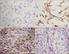
We isolated myoblasts from gastrocnemius muscles, and the cells were characterized by immunocytochemical staining. The cultured myoblasts were spindle shaped, and typical myoblast characteristics, as determined by Pax7 staining (Fig. 1). These cells, with potential differentiation capability based on MHC and SMA staining, were used in subsequent experiments.
Effects of autologous myoblast beads on CMAPs
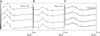
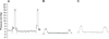
Three months after injuring the anal sphincter, the median amplitude of the CMAPs were significantly decreased compared with that measured before sphincter injury (1.22 mV vs. 3.00 mV, P = 0.04) (Table 1). Representative traces of CMAP are shown in Fig. 2. In the untreated sphincter injury group, there were no differences in the area of the CMAP between 3 months after sphincter injury and before injury (2.84 mV vs. 3.45 mV, P = 0.69). Meanwhile, in the autologous myoblast bead-treated group, the amplitude and area of the CMAP were not significantly different to those in the untreated group based on ANCOVA adjusted for baseline values.
Anal pressure
Three months after injuring the anal sphincter, both resting pressure and squeezing pressure were significantly decreased compared with the values measured before injury (respectively, 1.80 mmHg vs. 3.50 mmHg, P = 0.04; 4.02 mmHg vs. 9.00 mmHg, P = 0.04) (Table 1). Representative traces of anal pressure are shown in Fig. 3. Resting pressure and squeezing pressure were higher in the autologous myoblast bead-treated group compared with untreated injured group, although not significantly based on ANCOVA adjusted for baseline values.
Histologic findings
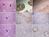
The external anal sphincter is composed of striated muscle while the internal anal sphincter shows characteristics of smooth muscle (Fig. 4A). The uninjured normal anal sphincter composed of smooth and skeletal muscle stained positively for both MHC and SMA (Fig. 4B, C). The injured anal sphincter showed damage to the muscle fibers with intermysial fibrosis and focal interstitial inflammatory cell infiltration (Fig. 4D). In addition, immunostaining showed atrophied striated and smooth muscle and loss of them (Fig. 4E, F). The anal sphincter of dogs treated with the autologous myoblast beads had a hypercellular inflammatory infiltrate that was partially surrounded by fibrous connective tissue (Fig. 4G), but the tissue showed no MHC staining and weak SMA staining (Fig. 4H, I). This inflammatory infiltrate was heterogeneous, consisting mainly of multinucleated giant cells containing foreign material in their cytoplasm, foamy macrophages and some lymphocytes; these findings are consistent with a foreign body reaction.
DISCUSSION
To our knowledge, this is the first study to examine whether a bioengineering technique could improve fecal incontinence. Unfortunately, the results did not support the use of porous polycaprolactone beads containing autologous myoblasts as a tool to improve sphincter function in a dog model of fecal incontinence. This is because the injected myoblasts did not integrate within the injured sphincter or play a functional role within the bioscaffold, even though we used porous beads that were previously found to stimulate the smooth muscle cell differentiation [19].
We consider that bioengineering techniques are necessary for patients with incontinence associated with large structural deformities. To date, several types of cells have been proposed for the regeneration of the anal sphincter muscle, including muscle-derived stem cells [12], bone marrow-derived mesenchymal stem cells [11], muscle progenitor cells [15], and myoblasts [23]. In the present study, we found limited differentiation of the myoblasts on histologic analysis, while a marked foreign body reaction was apparent around the site of injury. From these results, we consider that the pore size of the beads (25 to 50 µm) is too small to allow the entry of myoblasts, preventing integration of the cells and beads, even though the porous polycaprolactone beads containing autologous myoblasts are immune-compatible, nonmigratory, durable, and efficacious in other settings. The unexpected outcomes may also be related to an adverse immune response or granuloma formation following injection of an agent [8,24], but no foreign body reactions were reported in a Cochrane review [25]. In this study, the beads (200 to 300 µm in diameter) were larger than the maximum size of particles that can be ingested by macrophages. Clearly, the physiologic effects of foreign body reaction following injections of an agent, similar to that observed here, should be examined in animal models because no clinical studies have investigated the distribution of injected cells, or whether they remain viable. Another issue that needs to be evaluated in the context of injections of porous beads is whether the injected cells can differentiate into myofibers with contractile ability and can undergo reinnervation for functional muscle regeneration [12]. No studies have focused on tissue reinnervation in the field of muscle engineering, although some animal studies have indicated that muscle reinnervation may be possible by electrical stimulation, or the addition of Schwann cells [26,27].
In this study, the dog model was suitable and reliable for studying the effects of a potential treatment modality for fecal incontinence that could provide a greater step towards clinical trials than would studies in rats [11-14] or rabbits [15]. Indeed, a large animal model is needed to examine the specific effects on the anal sphincter, as the anal sphincter in rats is only a few millimeters thick and is almost impossible to isolate in vivo [28]. In this study using dogs, we were able to perform functional studies, including measurement of CMAPs of pudendal nerve. Thus, the dog model offers several advantages over rat and rabbit models, although it is limited by cost and convenience, which means rat models are often preferred over larger animals for studying novel treatments. Some functional studies in rats measuring in vitro contractility [11,12], and electromyography or manometry in vivo [13-15] have been reported quite recently. To our knowledge, ours is the first in vivo functional study using manometry and measurement of CMAPs in dogs. CMAP is considered to be a functional indicator of muscle function, although the amplitude of CMAPs is related to the diameter of the closest muscle fibers and the density of the muscle fibers comprising the motor unit closest to the electrode [29,30]. Therefore, the amplitude of CMAPs could be used as a marker of fecal incontinence in dogs.
In adult vertebrates, myogenesis is mainly sustained by a subpopulation of myogenic cells known as satellite cells, which are activated into myoblasts in response to stimuli, particularly tissue injury. Cultured myoblasts can also be integrated into the host muscle. The main advantages of autologous transplantation are the avoidance of tissue rejection, and the absence of ethical concerns, unlike stem cell therapy. Therefore, we used autologous myoblasts for the regeneration of the anal sphincter, as in earlier studies [16]. Further studies are needed to confirm whether the injection of porous polycaprolactone beads containing autologous myoblasts can stimulate muscle regeneration.
Our study has some limitations to discuss. First, the absence of statistical significance could be due to type 2 error, as there were only five dogs in each group, although some trends were evident. Second, we did not label the cells to enable cell tracking, a limitation of many studies of cell therapy. Further studies are planned to evaluate cell survival, distribution, differentiation, integration, and functional contributions of the cells using appropriate tracking methods. Third, CMAPs or anal manometry in the present study is not used as a stratifying tool for the severity of fecal incontinence in human, and was based on practical constraints of animal study that is not reliable with subjective tool of gas, liquid or solid stool incontinence.
Until recently, no definitive bioengineering approaches have been developed for fecal incontinence, as effective treatments require a reliable cell source, tissue vascularization, tissue function enhancement, biomaterial development, and tissue innervation. Although this study did not confirm that the injection of porous polycaprolactone beads containing autologous myoblasts can be used as a tool to improve sphincter function, we found that the dog model was suitable and reliable for studying the effects of a potential treatment modality for fecal incontinence.

 XML Download