

 ePub
ePub Citation
 Print
Print


INTRODUCTION
Hepatocytes are highly differentiated cells that perform many complex functions. There has been considerable interest in the control of growth and differentiation of hepatocytes in vitro; however, cultures of functional, differentiated adult hepatocytes have proved difficult to establish. Early attempts to culture adult hepatocytes invariably led to either overgrowth of contaminating cell types or to the dedifferentiation of the cultured hepatocytes [1-4]. More recently, several techniques have been explored to establish long-term cultures of functional hepatocytes. These include cultures 1) in arginine-free media, 2) on floating collagen membranes, 3) on various types of extracellular matrix materials, 4) together with other liver cell types, and 5) in the presence of dimethyl sulfoxide [5-9]. In each system, liver-specific functions have been maintained for periods ranging from 2 to 7 weeks. Although the use of various components of the extracellular matrix has been explored in hepatocyte culture, few attempts have been made to induce the formation of their rather specialized polarity by manipulating the extracellular matrix configuration.
Hepatocytes are epithelial cells with distinct apical (bile canalicular) and basal (sinusoidal) surfaces that serve different functions. Typically, hepatocytes have been cultured in systems that allowed cell attachment on one surface and medium on the opposite surface [1,2]. There are many reports of rat hepatocytes cultured in a collagen gel sandwich. However, studies of a similar culture system for mouse hepatocytes are scarce [2]. In this report, we describe the effects of sandwiching mouse hepatocytes between two layers of collagenous matrix, thereby providing an environment that more closely resembles in vivo geometry.
METHODS
Cell culture
Hepatocytes were isolated from 3-month-old adult C57/B6 mice (Charles River Laboratories, Boston, MA, USA) weighing 20 to 30 g using a modified version of a previously described two-step collagenase perfusion procedure in which primary mouse hepatocytes were sandwiched between two layers of collagen to maintain the stability of their liver-specific functions. Tissue culture dishes were coated with 0.5 mL of a mixed solution containing parts of rat-tail collagen (1.1 mg/mL in mM HCl) and 10 Dulbecco's modified Eagle Medium (DMEM) and incubated for 1 hour at 37℃ to form a collagen gel. After gelation, one million hepatocytes (12.5 × 10³ cells/cm2) were seeded in 2 mL hepatocyte culture medium and incubated in 90% air/10% CO2 at 37℃. To achieve uniform densities, the substrates were shaken every 15 minutes for the first hour after cell seeding. The following day, the culture medium was removed and a second collagen gel layer was overlaid on the hepatocytes and incubated for 1 hour at 37℃. After gelation, 2 mL of hepatocyte culture medium was applied. The culture medium was changed daily. The hepatocyte culture medium consisted of DMEM supplemented with 10% fetal bovine serum (Life Technologies Inc., Gaithersburg, MD, USA) ng/mL glucagon (Bedford Laboratories, Bedford, OH, USA), 7.5 g/mL hydrocortisone (Pharmacia Co., Kalamazoo, MI, USA), 0.5 U/mL insulin (Eli Lilly, Indianapolis, IN, USA), 20 ng/mL epidermal growth factor (Sigma Aldrich Co., St. Louis, MO, USA), 200 U/mL penicillin, and 200 g/mL streptomycin (Life Technologies Inc.).
RNA isolation
Total RNA was isolated from cultured mouse hepatocytes using TRIzol reagent with the RNeasy kit according to the manufacturer's protocol. Total cellular levels were measured on a spectrophotometer, and the quality of each RNA preparation was determined with a bioanalyzer. Extracted RNA was stored at -80℃ [3].
Reverse transcription-polymerase chain reaction (RT-PCR) for mouse primary hepatocytes
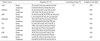
All quantitative PCRs were prepared using SYBR PCR supermix with the synthesized first-strand cDNA and specific primer pairs and performed using an ABI-PRISM 7500 Fast Detection System (Applied Biosystems, Foster City, CA, USA). The thermal cycling conditions were 10 minutes at 95oC, then 40 cycles of 95℃ for 30 seconds, 58℃ for 30 seconds, and 72℃ for 30 seconds. Expression of hepatic genes and that of the housekeeping gene, glyceraldehyde-3-phosphate dehydrogenase (GAPDH), were measured. Reaction conditions and primer sets are displayed in Table 1 [10].
RESULTS
Collagen gel sandwich for mouse primary hepatocyte culture
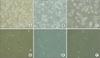
After 24 hours of culture, the primary mouse hepatocytes grown on the collagen gel sandwich (Fig. 1A) had a much clearer cellular border and more bile canaliculi than did those cultured using a single collagen coating (Fig. 1D). After 5 days, the mouse hepatocytes cultured on the collagen gel sandwich (Fig. 1B) maintained their cellular border and bile canaliculi while the cells grown using the collagen coating did not (Fig. 1E). The results after 10 days of culture were same as those at 5 days (Fig. 1C, F).
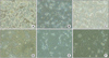
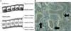
The differences between the collagen gel sandwich and the single layer systems were striking in the higher density (1 × 10⁶, Fig. 2) culture dishes. After 24 hours of culture, the mouse primary hepatocytes cultured using the sandwich system (Fig. 2A) had a considerably more distinct cellular border and a higher number of bile canaliculi than did those grown using the single collagen coating (Fig. 2D). After 5 and 10 days, the cellular border and bile canaliculi were maintained in the collagen gel sandwich system (Fig. 2B, C), but not in the collagen-coated dish (Fig. 2E, F). Furthermore, high power magnification of the culture dishes containing hepatocytes grown using the collagen gel sandwich (5 × 10⁴ cells/35 mm2 well, ×200; Fig. 3A) revealed clear apical and basal hepatocyte surfaces and an area corresponding to the space of Disse (Fig. 3B).
RT-PCR for mouse primary hepatocytes cultured using a collagen gel sandwich
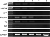
The results of RT-PCR targeting alpha-1-antitrypsin (AAT), hepatocyte nuclear factor 4 alpha (HNF4A), alpha-fetoprotein, albumin, tryptophan oxygenase (TO), the tyrosine aminotransferase (TAT) gene, glucose-6-phosphatase (G6P), and GAPDH differed between mouse primary hepatocytes grown on a single collagen layer and those grown using the collagen gel sandwich system (Fig. 4). The expression of AFP, TAT, and G6P disappeared in both mouse hepatocytes cultured in a collagen gel dish and those grown on a collagen-coated dish for 5 and 10 days. However, the expression of AAT, HNF4A, albumin, and TO was detected in mouse hepatocytes cultured on collagen gel after 5 and 10 days, but not in mouse hepatocytes grown on the collagen-coated dish (Fig. 4).
DISCUSSION
Primary hepatocytes have been used extensively to study hepatic uptake and metabolism [2]. The liver-specific functions of primary hepatocytes including albumin secretion, urea synthesis, CYP3A4 expression, and expression of tight junction-associated proteins have commonly been used in drug development [3]. Previous reports have demonstrated the influence that culture conditions including the type of culture media, media supplements, extracellular matrix and confluency have on cell morphology and bile canalicular network formation. It is known that normal hepatocytes are epithelial cells with distinct apical (bile canalicular) and basal (sinusoidal) surfaces that serve different functions. For example, bile acids are excreted into the bile duct by traversing the apical surface, whereas albumin is secreted into the circulation by traversing the basal surface. Unlike the classical epithelium, as typified by intestinal absorptive cells, hepatocytes have a belt of apical surface dividing two basolateral surfaces that are in contact with the extracellular matrix. Typically, hepatocytes have been cultured in systems that allowed cell attachment on one surface and medium on the opposite surface. The extracellular matrix has been found to play an important role in maintaining hepatocyte functions [2,13].
Along with the improvement in enzyme digested hepatocyte isolation techniques in recent years, hepatocyte culture has been researched extensively [1-3]. Numerous culture methods have been used or are under investigation to facilitate the functional cell growth of primary hepatocytes in vitro. Polystyrene culture plates or dishes are most commonly used for cultures of HepG2 cells or primary human and animal hepatocytes [7]. However, due to the complexity of the liver structure and function as well as the specificity of hepatocytes in vivo, isolating hepatocytes in vitro requires very strict conditions. The monolayer attachment culture model is not suitable for hepatocytes isolated in vivo as it only meets general experimental requirements. In order to overcome this difficulty, an enormous amount of research has been carried out to improve media, supplements, growth factors, matrix composition, and techniques for monolayer hepatocyte culture. However, this method requires high density, high activity, and long-term hepatocyte culture, which are not yet possible. Therefore, the exploration of hepatocyte culture with high activity has become an important subject in this research field [1-4].
In this report, we describe the effects of sandwiching hepatocytes between two layers of collagenous matrix, thereby providing an environment that more closely resembles the in vivo geometry. The sandwich-cultured hepatocyte system is composed of two compartments: cytosol and canalicular lumen. The tight junctional complex is the diffusional barrier between the canalicular lumen and the extracellular space [11,12]. Many studies have indicated that primary rat hepatocytes cultured between two layers of collagen (sandwich configuration) maintain normal morphology, form extensive canalicular networks, and exhibit sustained liver functions [12]. In this experiment, primary mouse hepatocytes cultured using the collagen gel sandwich maintained their cellular border and bile canaliculi better than did cells grown on a single collagen coating after 24 hours, 5, and 10 days of culture, indicating that mouse hepatocytes can maintain their growth and specific functions when grown in a collagen gel sandwich. High-power magnification of culture dishes containing the collagen gel sandwich clearly showed that the cells exhibited apical and basal surfaces and the space of Disse. RT-PCR analysis of AAT, HNF4A, alphafetoprotein, albumin, TO, the TAT gene, G6P, and GAPDH expression in mouse primary hepatocytes grown in the collagen gel sandwich indicated that the collagen gel sandwich culture system is superior to the single-layer collagen culture system with regard to the maintenance of hepatocyte form and function similar to that in vivo.
Long-term (more than 24 hours) sandwich-cultured hepatocytes represent a potential in vitro model with which to study biliary excretion. Previous work has demonstrated that maintenance of hepatocytes in a collagen-sandwich configuration prolongs cell viability and preserves liver-specific protein synthesis. Further studies showed that long-term sandwich-cultured hepatocytes reestablish a structurally and functionally normal bile canalicular network and better maintenance of drug uptake and enzyme-induction potential [11,12].
These reports in conjunction with our findings illustrate that the collagen gel sandwich configuration could enable mouse hepatocyte culture and growth with high density and high activity. The advantage of this culture method is that the structure of the cultured hepatocytes is similar to that of in vivo and that it facilitates high cell density and extensive connections between mouse hepatocytes and other cells. Therefore, the collagen gel sandwich method for mouse hepatocyte culture may serve as a useful means of studying hepatic uptake and metabolism of mouse adult hepatocytes.
In summary, primary mouse hepatocytes cultured between layers of collagen or extracellular matrix in a sandwich configuration are commonly employed as an in vivo model to examine the hepatic disposition and metabolism of compounds and to determine mechanisms of hepatotoxicity. When cultured in a conventional monolayer, cells dedifferentiate over time and rapidly lose their hepatocytic functions. However, when cultured in a sandwich configuration, mouse hepatocytes continue to synthesize and secrete hepatocyte-specific compounds such as albumin, transferrin, and fibrinogen. Thus, the collagen gel sandwich configuration for mouse hepatocyte culture method is suitable for adult mouse hepatocytes.
 XML Download