

 ePub
ePub Citation
 Print
Print


INTRODUCTION
The incidence of thyroid cancer has increased continuously worldwide [1,2]. Papillary thyroid carcinoma (PTC) is the most common thyroid malignancy, accounting for up to 80% of primary thyroid cancers [3]. Cervical lymph node (LN) metastasis frequently occurs in patients with PTC, and up to 90% of patients with new thyroid malignancies are found to have LN metastases [4]. Since LN metastasis is a risk factor for recurrence [5]. The detection of such metastases prior to or during initial surgery may affect recurrence and patients survival.
Although ultrasonography has been shown to be the most useful tool for preoperatively detecting cervical LN metastases, its accuracy has been found to vary widely, from 40% to 90%, and to be operator-dependent [6]. Use of a combinatioin of ultrasonography and a second method, such as intraoperative sentinel LN biopsy (SLNB), may identify LN metastases more [7-9].
SLNB is the gold standard for detecting LN metastases in patients with melanoma and breast cancer. This technique has also been utilized in patients with thyroid cancer, using vital blue dye [10], or a radioisotope with favorable results observed in patients with PTC [11,12]. A complementary method, preoperative lymphoscintigraphy defines the drainage basins and SLN locations of individual tumors [13].
However, because LN drainage in the head and neck is unpredictable and ambiguous, preoperative lymphoscintigraphic SLN mapping is important for tailoring the surgical field in individual patients [14]. Since planar scintigraphy did not provide the precise location of SLN, due to poor anatomic information, single photon emission computed tomography/computed tomography (SPECT/CT) has been used to preoperatively map SLN in patients with head, neck and gynecologic cancer. We have, therefore, investigated the usefulness and accuracy of SLNB, as detected by preoperative SPECT/CT with technetium-99m phytate, of locoregional LN metastases in patients with PTC. To our knowledge, this is the first report on the use of SPECT/CT for localizing SLN in PTC.
METHODS
Patients
From July 2010 to March 2011, 39 patients with differentiated PTC underwent SLNB of the central and lateral neck compartments in our institution. All patients had been preoperatively diagnosed with PTC by fine needle aspiration (FNA) biopsy and had risk factors for recurrence or required intraoperative LN sampling for suspected LN metastases on preoperative imaging. No patient had a history of either thyroid or neck surgery. Patients with other types of thyroid malignancies were excluded. The study was approved by the Institutional Review Board of our institution and written informed consent was obtained from each patient.
Scintigraphic technique
Twenty MBq 99mTc phytate was injected into each tumor, using a 26-gauge syringe. Early lymphoscintigraphy and SPECT/CT were performed 10 minutes later. Delayed lymphoscintigraphy and SPECT/CT were performed, 2 hours after injection. Surgery was performed 2 to 4 hours after injection. Anterior planar images were obtained during lymphoscintigraphy. SPECT/CT emission/transmission was performed using a hybrid system consisting of a dual-head gamma camera with a low-dose X-ray tube installed in its gantry (Infinia Hawkeye 4 SPECT-CT, GE Healthcare, Waukesha, WI, USA). This system allows both transmission and emission acquisitions without changing the patient's position. SPECT acquisition parameters for SLN detection included a matrix size of 128 × 128, 180 degree in anterior L-moderotation, and a 4 degree angle step with 25s time frame. Transmission data of the patient were corrected and reconstructed using the filtered back-projection to produce cross-sectional attenuation of the imaged tissue. The SPECT and CT images were fused on the Xeleris Function Imaging Workstation version 2.1507. The hottest node found positive on both preoperative lymphoscintigraphy and SPECT/CT was considered the sentinel LN, as previously reported [15].
SLN identification at surgery
Under general anesthesia, a standard, transverse, low-collar skin incision was made, and the myocutaneous flap was lifted. A fascial incision was made between the strap musculature and the sternocleidomastoid muscle, exposing the ipsilateral jugular vein. The SLN was harvested using the intraoperative hand-held probe (Neoprobe 2000, Johnson & Johnson Medical, Hamburg, Germany) and sent for both frozen and permanent sections. Total thyroidectomy was completed subsequently and all LNs of the central compartment were dissected. Modified radical neck dissection (mRND) was performed only in patients with positive LNs, and the nodes were labeled and sent for permanent histological section. All patients underwent compartment dissection at cervical levels II, III and IV.
The radioactivity counts of the lymphatic basin were assessed before and after excision of each node, and the radioactivity of each excised node was recorded after excision. Any node that had a count of at least 10% of the radioactivity of the hottest node was excised.
Data analysis
Pathologic specimens were stained with hematoxylin and eosin and were observed under light microscopy. The sensitivity, specificity, accuracy, and positive predictive value (PPV) and negative predictive value (NPV) of SLN biopsies, were calculated. Other variables were investigated using t-test and chi-square tests. All statistical calculations were performed using SPSS ver. 13.0 (SPSS Inc., Chicago, IL, USA). Postsurgical morbidity information was obtained from follow-up clinical and laboratory data.
RESULTS
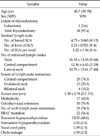
We assessed 39 patients (9 males and 30 females), of mean age 45 years (range, 30 to 70 years). Mean (±standard deviation) tumor size was 1.30 ± 0.73 cm. Bilateral jugular LN metastases were found in 4 patients, and unilateral jugular LN metastases in 13. The demographic and clinical characteristics of these patients, as well as their treatments and morbidities, are summarized in Table 1.
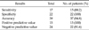
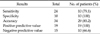
A SLN was detected in 38 patients (97.4%). The mean number of SLNs per patients was 4.73 ± 3.06 (range, 0 to 12), the mean number of lateral compartments, in which SLN was detected, was 2.15 ± 0.921 (range, 0 to 4), and the mean number of SLNs per lateral compartment was 2.22 ± 1.36 (range, 0 to 6). The sensitivity, specificity, accuracy, PPV and NPV of SLNB for lateral LN metastasis were 88.2%, 100%, 94.8%, 100%, and 91.6%, respectively (Table 2). The sensitivity, accuracy and NPV for lateral LN metastasis were higher than those for central LN metastasis (Table 3).
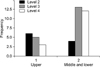
The association between the tumor sites and the pattern of LN metastasis in the ipsilateral compartment was not statistically significant (P = 0.055) (Fig. 1). In the patient with the skip metastasis, the primary tumor was located in the upper portion of the thyroid gland.
When we calculated the concordance rate based on cervical levels between suspicious LNs on US and SPECT/CT, the mean concordance rate was 27.9%.
When we compared preoperative US-guided FNA with intraoperative SLNB in 11 patients, the sensitivity of SLNB was higher than that of preoperative FNA at lateral neck (88.2% vs. 33.3%) and central LN metastases in 7 patients among them were diagnosed with SLNB only.
DISCUSSION
PTC is a representative histological type of thyroid cancer. It shows a mild behavior and has an excellent prognosis. However, LN metastases are frequently detected, primarily in cervical LN. Cervical LN metastases have been associated with a higher incidence of recurrence [16,17]. Although radioiodine treatment may reduce recurrence, 131I ablation was effective in only 10% of high-risk patients according to the AMES (age, metastasis to distant sites, extrathyroidal invasion, size) classification, and was not effective in 30% of low-risk patients [18].
Ultrasonography is useful in the preoperative assessment of cervical LN metastasis in patients with PTC [6]. However, a diagnosis of LN metastasis is difficult, because most patients with PTC have multiple, very small nodes <5 mm in diameter and metastatic LNs may show no change on serial imaging over several years [3]. Therefore the accuracy of ultrasonography in detecting cervical LN metastasis is not very high. The accuracy of loss of fatty hilum, round shape, calcification, cystic change or abnormal vascularity has been reported to range from 51% to 77% [19]. Ultrasonography may be complemented by measuring Tg in FNA (FNA-Tg) or by LN sampling during surgery. However, false positive or false negative FNA-Tg results have been reported, although their frequencies were low, and FNA-Tg cannot be performed in patients with obstruction of the carotid artery [20]. Moreover, it may be impossible to dissect LNs targeted during intraoperative sampling, because of the necessity to perform blunt dissection without anatomic landmarks. Therefore, in these patients, SLNB may accurately diagnose LN metastases.
To date, indication for SLNB have not been established. The recurrence rate in cervical LNs has been reported to range from 5.8% to 10.7% [6,21,22], with risk factors for recurrence including age, tumor size, extrathyroidal extension, LN metastasis and sex, et al. [6,21]. For example, recurrence rates were reported to be higher in patients <30 years of age and patients with larger sized tumors, cervical LN metastasis, local invasion [21]. Another study reported that risk factors for recurrence were age >54 years, LN metastasis and tumor size >4 cm [6].
Prophylactic mRND has been recommended to prevent recurrence in cervical LNs [6,23]. For example, patients with LN metastasis or, capsular invasion and woman >60 years of age were reported to benefit from mRND [23]. Similarly, prophylactic mRND may be beneficial in patients with two or more of the following characteristics: male gender, age ≥55 years, maximal tumor diameter >3 cm, and massive extrahyroid extension of the tumor [6]. Due to the high morbidity rates associated with mRND, however, the gold standard of lateral LN management is not prophylactic, but therapeutic mRND.
Therefore selective SLNB may determine whether therapeutic mRND should be utilized in patients <30 or >54 years of age, with tumor size >3 cm in diameter, or with local invasioin, or to reduce false positive or false negative FNA cytology or FNA-Tg results.
We assessed the usefulness and accuracy of SLNB in patients with risk factors for recurrence or requiring intraoperative LN sampling for suspected LN metastases on preoperative SPECT/CT. We found that the sensitivity, specificity, accuracy, and PPV of SLNB for lateral and central LN metastases were comparable to results from previous reports that did not utilize SPECT/CT, with sensitivity ranged from 77.7% to 91% and accuracies from 90% to 95.3% [24,25]. In addition, the high resolution and anatomic information of SLN provided by SPECT/CT made intraoperative gamma probing easier, compared with information provided by palpation and inspection [15].
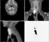
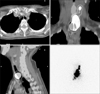
In addition, SPECT/CT may also show pathway of lymph basins. We observed one patients with a lymph basin extending from the right jugular LN at cervical level II to the right retropharyngeal LN (Fig. 2), and another with a basin extending from the right paratracheal to the right high mediastinal LN (Fig. 3). However, the distribution of SLNs was not similar to that of metastatic LNs in the lateral neck. The ipsilateral distribution of SLNs showed the highest frequency at level IV, whereas the distribution of metastatic LNs showed the highest frequency at level III.
Generally, lymphatic metastasis from differentiated PTC occurs in a stepwise manner from the LNs in the tracheoesophageal groove to the LNs in the jugular chain, including the supraclavicular fossa [26]. Following the initial metastases in the central compartment, LN metastases were observed in the deep inferior and lateral cervical nodes, with no relation with tumor location [27]. We also found that the pattern of LN metastasis is not related to the primary tumor site. However, a skip metastasis, defined as lateral without central LN metastasis and observed in 7.7% to 15.3% of patients [28-30], was observed in only one patients with PTC in the upper portion of thyroid gland. Therefore, the relationship between tumor location and the pattern of LN metastasis requires further investigation.
Although SLNB is useful to identify the LN metastasis, it has two demerits. Unnecessary lateral LN dissection can be performed in spite of subclinical LN metastases, which are not related with recurrence, because the frequency of occult LN metastases of papillary thyroid cancer is 40% to 90% [10]. In addition, SLNB takes long surgical time because two or more SLN are identified in most of our patients (Table 1) dut to the rich lymphatic system of the thyroid gland. Therefore, routine SLNB may result in longer surgical time and hospitalization with increased surgical morbidity.
In conclusion, SLNB using SPECT/CT with technetinum-99m may be useful in accurately assessing LN stage during surgery patients with risk factors for recurrence or those requiring intraoperative LN sampling for suspected LN metastases on preoperative imaging. However, further studies are required for appropriate method of SLNB, by which the distribution of SLNs can be closer to those of metastatic LNs for the sake of avoiding unnecessary dissection in 67% of the patients without LN metastasis.
 XML Download