

 ePub
ePub Citation
 Print
Print


INTRODUCTION
Intrahepatic duct (IHD) stone is a common disease in Southeast Asia and it is occasionally associated with severe stricture of intrahepatic biliary trees. Stone removal and maintenance of adequate biliary flow are the bases of its treatment. Although there are various treatment modalities for IHD stones [1-4], hepatectomy seems to be the definitive procedure as it can remove IHD stones and stricted bile duct, simultaneously [4,5]. There have been difficulties in laparoscopic liver resection for IHD stones because of adhesion of adjacent tissue or distorted anatomy resulting from recurrent inflammation. Improvements of laparoscopic instruments and increasing experience with laparoscopic surgery have made laparoscopic hepatectomy a new procedure for various liver lesions including IHD stone [6].
In this study, we analyzed our experience with laparoscopic surgery for left IHD stone to evaluate its feasibility.
METHODS
Patients
From January 2009 to June 2011, a total of 26 consecutive patients underwent total laparoscopic left hemihepatectomy for left IHD stones at Gyeongsang National University Hospital. The indications for performing laparoscopic left hepatectomy were 1) impacted stone in the left liver, 2) stones associated with stricture or dilatation of the left IHD, and 3) left IHD stones associated with parenchymal atrophic changes. Before operation, all patients had complete medical evaluation, including liver function, renal function, electrocardiogram and chest X-ray. Preoperative ultrasonography or abdominal computed tomography (CT) was performed in order to identify the distribution of stones and changes in the bile duct trees. In cases where common bile duct (CBD) stones were found in preoperative image, we performed endoscopic retrograde cholangiopancreatography (ERCP) and endoscopic sphincterotomy (EST) and removed the CBD stones. All of the patients underwent abdominal CT or ultrasonography at 7 days after operation for detection of any remnant stones. We retrospectively reviewed the clinical outcomes and the stone clearance rates of the 26 patients in this study. Informed consent was obtained from all the patients before surgery.
Operative technique
Under general anesthesia, the patients were placed in the supine position with a 30° reverse Trendelenburg position. Both legs were separated at about 60 degrees. The position was changed according to resection plane.
A pneumoperitoneum was established through a 10-mm umbilical port, and this was maintained below 12 mmHg.
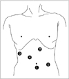
The second 10-mm trocar was placed on the left side of the upper midline to facilitate liver retraction. The third 5-mm trocar was inserted at the right lateral side below the right costal line. The forth 12-mm trocar was inserted between the second and the third trocar about 3 cm caudal to the third trocar. The fifth 5-mm trocar was placed on the left lateral side above the umbilical line (Fig. 1).
Trocars were basically placed at the above-described positions, subject to slight variations according to intra-abdominal conditions of the patient. It is better for the operator's trocar to be placed close to the resection line and for the resection line to be positioned in front of the operator. In cases of total laparoscopic left hemihepatectomy, the operator always stood at the right side of the patient to perform the operation and used the third and the forth trocar. The cameraman sat between the patient's legs and the operator was able to secure an unobstructed space with a stable field of vision throughout the long hours of operation. The assistant used 2 trocars on the left side and performed proper liver retraction and suction of bleeding sites as in open hepatectomy to enable the operator to have a full field of vision.
Cholecystectomy was initially performed in the usual manner. Before hepatic parenchymal dissection, dissection at the porta hepatis was performed and the left hepatic artery and left portal vein were isolated and ligated individually. Then, for mobilization of the left liver, the ligaments around the left liver, including the left triangular ligament and the falciform ligament, were dissected until the left hepatic vein was exposed.
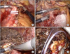
The superficial hepatic parenchyma was transected using ultrasonic shears (Harmonic scalpel, Ethicon, Cincinnati, OH, USA; Sonosurg, Olympus, Tokyo, Japan) and the deeper portion of the parenchyma was dissected using a laparoscopic cavitron ultrasonic surgical aspirator (CUSA, Valleylab, Boulder, CO, USA). The left hepatic vein was divided with a vascular endo-GIA. After dissection of the liver parenchyma, the left hepatic duct was isolated. In the preoperative study, if the stones were presumed to be located close to the resection line, then the duct was divided and the stones were extracted. Intraoperative choledochoscopy was performed to confirm residual stone. Then the duct was closed with intracorporeal sutures (Fig. 2). In the preoperative image, if stones were located far from the resection line, then the duct was transected with an endo-GIA. Once the resected specimen was completely divided, it was inserted into a vinyl bag and left in the right-side intra-abdomen.
After careful hemostasis, a fibrin glue sealant (Greenplast, Green Cross Co., Seoul, Korea) was applied to the raw surface. After irrigating the surgical field, a silastic drain was inserted. Finally, a surgical specimen was extracted though a small incision that was created by extending the wound at the umbilical port site and the wound was closed in layers.
RESULTS
Patient characteristics and perioperative outcome
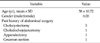
The mean age of the patients was 58 ± 10.72. The patients were 6 men (23.1%) and 20 women (76.9%). Six patients had a history of abdominal surgery including cholecystectomy (n = 3), choledochojejunostomy (n = 1), appendectomy (n = 1), and cesarean section (n = 1). Total laparoscopic left hemihepatectomy was performed successfully in all 26 patients (Table 1).
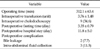
The mean operation time was 312.1 ± 63.4 minutes. Intraoperative transfusion was needed for 23 patients (88.5%). The causes of transfusion were bleeding and anesthetic principles of our institution (hemoglobin < 10 g/dL during operation, intraoperative central venous pressure was lower than initial central venous pressure). The mean duration of postoperative hospital stay was 11.8 ± 5.0 days. Intraoperative choledochoscopy was performed in 9 cases (34.6%). There were 2 cases of postoperative bile leakage and 3 cases of intra-abdominal fluid collection that needed additional management including percutaneous drainage and antibiotics (Table 2).
Cholangiocarcinoma was combined in 1 case. In this case, cholangiocarcinoma was suspected in the surgical specimen and laparoscopic lymph node dissection was performed simultaneously. The resection margin of the bile duct was not involved by the tumor. The patient is alive without recurrence after 17 months.
Outcome of stone clearance
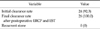
Remnant stones, as confirmed by abdominal ultrasonography or abdominal CT at 7 days postoperation were detected in 2 patients. The initial success rate of stone clearance was 92.3% (24 of 26). The remnant stones were located in the CBD in both cases and were removed by ERCP and EST. Therefore, the final success rate of stone clearance was 100% (26 of 26) (Table 3).
During a mean follow-up of 22 months (range, 7 to 36 months), there was no patient with recurrent stone.
DISCUSSION
IHD stones are a prevalent disease in Southeast Asia and the incidence in the Korean population has been reported to be 15% of all biliary tract stones, which is relatively higher than the data reported for Western populations [7].
Hepatectomy was considered the most effective and safe procedure with a high stone clearance rate, low morbidity and a low long-term stone recurrent rate. For treatment of IHD stones, hepatectomy is a safe and useful treatment that can remove stones and associated pathologic changes, including ductal stricture, microabscess, and fibrosis by a single operation [4,5].
Laparoscopic liver resection has developed more slowly than others because of the complex anatomy of liver, the technical difficulty, the risk of massive bleeding, air embolism and the relatively long learning curve [8,9]. However, with the accumulated experience of surgeons and the improvement of laparoscopic instruments, an increasing number of reports on laparoscopic liver resection for various hepatic lesions have been reported. However, there have been only a few reports on laparoscopic liver resection for IHD stones [10-13].
From 2008, laparoscopic procedures have been performed for IHD stones in Gyeongsang National University Hospital.
In the early period, 11 patients with IHD stones underwent laparoscopy-assisted hepatectomy. For mobilization of the left liver, the ligaments around the left liver were sharply dissected laparoscopically until the left hepatic vein was exposed. Then, an approximately 10 to 15 cm sized upper midline skin incision was created. Hepatic parenchymal transection was performed in the same manner as open technique. Based on these experiences, total laparoscopic liver resection was performed in the late period.
Most laparoscopic liver resection is performed using 4 to 7 trocars. Selecting the positions of the trocars is crucial for liver retraction to secure an accurate field of vision and for facilitating handling of laparoscopic instruments. We used 5 trocars (three 10-mm trocars and two 5-mm trocars) in all patients.
Laparoscopic liver resection for IHD stones is more technically demanding because of severe perihepatic adhesion and anatomic distortion resulting from the recurrent inflammation.
In addition, parenchymal transection is often difficult because of parenchymal fibrosis.
Difficulty of hemostasis is a major concern in laparoscopic liver resection and is a major cause of open conversion [6]. To avoid massive bleeding during parenchymal transection, the Pringle's maneuver has been used in some laparoscopic liver resections [9,14-16]. Pringle's maneuver was not used in our cases. Before parenchymal transection, the hepatic artery and left portal vein were isolated and ligated individually. Selective inflow occlusion is more technically demanding than total vascular occlusion in cases of IHD stones because of the peripheral adhesion or anatomic distortion resulting from recurrent inflammation. But it can prevent complications of ischemic reperfusion injury and gastrointestinal congestion [17,18]. Furthermore, selective inflow occlusion allows surgeons to the take time needed for meticulous dissection because it does not require fast liver transaction [19]. To prevent complications such as biliary fistula, parenchymal dissection should be more carefully performed because of parenchymal fibrosis and deformed intraheptaic biliary anatomy.
The stone clearance rate after open surgery has been reported to be 75 to 98% [4,20-22]. Since, by comparison, the stone clearance rate after laparoscopic surgery has been reported to be higher than 80%, there is no significant difference between the two groups [10-12]. In our study, the initial stone clearance rate was 92.3% and the final stone clearance rate was 100%. Our data showed a similar outcome for the stone clearance rate. The use of intraoperative choledochoscopy or intraoperative ultrasonography could further raise the stone clearance rate.
In conclusion, laparoscopic hepatectomy for IHD stones is compare to open hepatectomy in results of stone clearance rates. Therefore, laparoscopic surgery could be an effective treatment option for the management of IHD stones in selected patients.
 XML Download