

 ePub
ePub Citation
 Print
Print


INTRODUCTION
In adult general surgery setting, postoperative adhesive obstruction which needs for laparotomy is reported in small portion of patients. However, bowel obstruction after liver transplantation is an unusual complication. Intussusception is a rare cause of intestinal obstruction in adults compare to the pediatric population. In adults, some underlying cause such as a benign or malignant tumor is present at the leading point of the intussusception in about 90% cases. Therefore, surgery is needed for the treatment of intussusception in most cases. Post transplant lymphoproliferative disorder (PTLD) is a relatively common malignancy after transplantation and accounts for up to 10% of all solid organ transplant recipients. We present a rare case of intussusception associated with PTLD in adult liver transplant patient.
CASE REPORT
A fifty-year-old male underwent deceased donor liver transplantation in September 2003 due to liver cirrhosis, secondary to chronic hepatitis C infection. The donor was a 27 year-old male who died of head trauma. Both donor and recipient were positive for cytomegalovirus and Epstein-Barr virus (EBV) antibodies in the serum. Cold ischemia time was 5 hours 43 minutes. Daclizumab (anti-CD25 monoclonal antibody) 1 mg/kg was used for induction therapy. Tacrolimus and steroids were used as maintenance immunosuppressants. Steroids were tapered off 3 months after transplant. There was no episode of acute rejection or evidence of recurrent hepatitis C until the patient developed PTLD.
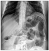
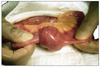
He was admitted to the hospital with acute abdominal pain 7 years after transplant. Simple abdominal X-ray showed multiple dilated small bowel loops with air fluid levels, which were resolved with nasogastric tube decompression within 24 hours. No further study or specific treatment was given, and the patient was discharged. The patient required re-admission 7 days later with symptoms and signs of complete intestinal obstruction. Simple abdominal X-ray showed findings similar to those on the previous admission (Fig. 1). Because this was the second episode within a short period of time, an emergency laparotomy was performed without further radiologic imaging, such as abdominal computed tomography (CT) scan. Operative finding revealed intussusception of the jejunum, approximately 300 cm distal from ligament of Treitz (Fig. 2). No abnormal lymphadenopathy was visualized in the mesentery of the small intestine. Simple resection of the small bowel was performed since no other pathology was found in the abdominal cavity. Post-operative recovery was uneventful, and the patient was discharged 4 days after surgery.
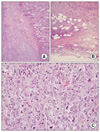
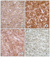
The surgical specimen showed a tan firm, well-circumscribed mass, measuring 4 × 3.9 cm, which occluded the lumen of the jejunum. Microscopically, the tumor extended from the mucosal surface through the muscularis layers and approached the serosal surface. The tumor cells were large, had high nuclear to cytoplasmic ratio, delicate chromatin, and exhibited prominent centroblastic features (Fig. 3). The tumor cells were positive for CD20, CD79a, CD10, and BCL-6 by immunohistochemistry (Fig. 4). They were negative for CD3, CD5, BCL-2, S-100 and keratin. Ki-67 index was high (greater than 90%). Fluorescent in situ hybridization analysis was positive for translocation between chromosomes 8 and 14 (IGH/MYC) in 10% of interphase nuclei, and negative for translocation between chromosomes 14 and 18. In situ hybridization demonstrated that the tumor was negative for EBV. Based upon these things, a diffuse large B-cell lymphoma of germinal center phenotype with MYC translocation was diagnosed.
A complete staging evaluation was performed, which confirmed stage IIAE disease with an international prognostic index score of 0. The patient received 6 cycles of chemotherapy consisting of rituximab (anti-CD20 monoclonal antibody) 375 mg/m2, cyclophosphamide 750 mg/m2, doxorubicin 50 mg/m2, vincristine 1.4 mg/m2 (capped at 2 mg) IV, and prednisone 100 mg orally daily for 5 days (R-CHOP) given every 14 days, followed by Neupogen (Amgen Inc., Thousand Oaks, CA, USA) growth factor support. The patient tolerated the treatment without any major adverse effects and achieved a complete remission.
DISCUSSION
Postoperative intra-abdominal adhesions represent the most common cause of intestinal obstruction in non-transplant operations, with a very small percentage (3.3%) requiring laparotomy [1]. Intestinal obstruction after liver transplant has not been extensively studied, but seems to be a rare complication. Blachar and Federle [2] reported a 1.2% incidence of intestinal obstruction in 4,001 liver transplantations performed during a 15-year period. Unusual causes such as internal hernias around the infrarenal arterial conduits or through the mesenteric defect for Roux-en-Y hepaticojejunostomy, diaphragmatic hernias, and intussusceptions are reported [3-5]. In pediatric liver transplant patients, there are a few reports describing intussusception associated with PTLD, but these are extremely rare in adult patients to the best of our knowledge. Clinical presentation of the intussusception is usually intestinal obstruction; however jaundice was reported when it occurred at the Roux limb or jejunojejunostomy which mimicked biliary obstruction [4,5]. In adults, most intussusception has a benign or malignant tumor as a leading point, which necessitates surgical resection [6]. In cases of incidental or asymptomatic intussusception in pediatric liver transplant patients, close follow-up without surgical intervention was reported as a possible treatment option [4,7]. In general, intestinal obstruction after liver transplant is rare; but unusual causes, other than adhesions, are more frequently reported than in non-transplant surgery. Laparotomy should be always considered as a treatment option.
PTLD is a relatively common malignancy after transplantation and occurs in up to 10% of all solid organ transplant recipients [8]. It is the most common cause of cancer-related mortality after solid organ transplantation in both children and adults. The incidence of PTLD after solid organ transplantation varies according to the type of graft and differs between children and adults. The incidence of PTLD in adult liver transplant recipients has been reported as 1 to 3%. EBV induced PTLD is well documented in the literatures and most cases of early PTLD, i.e., polymorphic lymphoid proliferations occurring within one year of transplant, are related to primary infection with EBV or reactivation of previously acquired EBV. In contrast, late PTLD (as in our case) presents with histologic features of non-Hodgkin lymphoma, most frequently diffuse large B-cell lymphoma; and the majority of cases are not associated with EBV infection. Treatment of early (polygonal) PTLD should include a reduction or discontinuation of immunosuppressive agents. Rituximab has been used either as a single agent or in combination with other treatment modalities in the treatment of patients with early and late PTLD after solid organ transplantation. Late (monoclonal) PTLD is generally unresponsive to a reduction in immunosuppression and should be treated with chemotherapy according to the underlying histologic lymphoma subtype [9,10].
This is a very rare case of intussusception, associated with PTLD, in an adult liver transplant patient. In the clinical setting of intestinal obstruction after liver transplant, this unusual cause of intestinal obstruction should be always considered.
 XML Download