

 ePub
ePub Citation
 Print
Print


INTRODUCTION
Riedel's thyroiditis is a chronic inflammatory disease of the thyroid gland characterized by invasive fibrosis that partially destroys the thyroid gland and extends into adjacent neck structures [1,2]. It was recognized in 1986 by Riedel [3], who described two patients with hard goiters and tracheal compression symptoms.
The diagnosis of Riedel's thyroiditis is clinically difficult because this form of thyroiditis can mimic malignant neoplasm or the fibrous variant of Hashimoto thyroiditis during preoperative physical, radiologic, and pathologic examination [4-6].
We describe here a rare case of Riedel's thyroiditis in an elderly patient and review the literature for its radiologic characteristics. Written informed consent was obtained from the patient for publication of this case report.
CASE REPORT
A 77-year-old female patient presented with general weakness, dysphagia and dyspnea. On physical examination, there was a firm, painless, large mass in the anterior neck and wheezing sounds were noted on auscultation.
A thyroid function test was abnormal, which showed hypothyroidism: T3 of 62.4 ng/dL (71 to 161 ng/dL), free T4 < 0.1 ng/dL (0.8 to 1.7 ng/dL), thyroid stimulating hormone (TSH) > 100 µIU/mL (0.86 to 4.69 µIU/mL), TSH receptor antibody (Ab) of 0.70 U/L (0 to 1.5 U/L), thyroglobulin antigen of 0.9 ng/mL (0.1 to 32.5 ng/mL), anti-thyroglobulin Ab of 1,359.0 IU/mL (10 to 124.2 IU/mL), and anti-thyroid peroxidase Ab > 600 IU/mL (5 to 13.5 IU/mL).
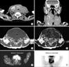
Imaging modalities including ultrasonography (US), computed tomography (CT), magnetic resonance imaging (MRI), and positron emission tomography (PET) scan were performed (Fig. 1). All showed a diffusely enlarged mass covering both thyroid lobes, extending to the infra-hyoid level and encircling the trachea and thyroid cartilage. This mass caused tracheal stenosis, but there was no evidence of tracheal invasion on MRI.
Despite the combination of these imaging modalities, the thyroid mass was not differentiated from a malignant neoplasm such as anaplastic thyroid carcinoma, or severe thyroiditis. Preoperative pathologic diagnostic procedure including fine-needle aspiration cytology (FNAC) was not performed because we have thought that FNAC might not be helpful for differentiating anaplastic thyroid cancer from severe thyroiditis and operation should be performed for relieving the compressive symptoms.
Operation was performed. First, the pyramidal lobe and isthmus were resected, and an intraoperative frozen section was performed due to the suspicion of anaplastic thyroid carcinoma. The results of the frozen section demonstrated that there were no malignant epithelial cells, and the possibility of Riedel's thyroiditis was suggested. Even though the benign result of the frozen section, bilateral total thyroidectomy was performed clearly without complications due to the purpose of relieving the compressive symptoms and tiny possibility of malignancy (total weight, 361 g).
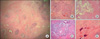
Hematoxylin and eosin staining showed extensive fibrosis and calcification with lymphoplasmacytic infiltration. Immunohistochemical stainings with various markers were performed for differential diagnosis with the following results: presence of apple green birefringence in Congo red staining, negative for acid-fast bacilli, and negative for calcitonin and carcinoembryonic antigen. Taken together, Riedel's thyroiditis was the final pathological diagnosis (Fig. 2).
The patient was discharged from the hospital 4 days after surgery without any complications such as bleeding or hypocalcemia. The patient has received routine check-ups and thyroid function tests along with thyroid hormone replacement (L-thyroxine 0.05 mg/day).
DISCUSSION
Riedel's thyroiditis is extremely rare. Although the etiologic mechanisms underlying Riedel's thyroiditis are unclear, the prevailing view is that it is part of a generalized fibro-inflammatory process that also involves other organs [7]. The main characteristics of Riedel's thyroiditis is invasive fibrosis that partially destroys the thyroid gland and extends into adjacent neck structures [1,2].
It is difficult characteristics for physicians to distinguish Riedel's thyroiditis from malignant neoplasms of the thyroid clinically because both clinical examination and imaging of Riedel's thyroiditis suggests malignancy [8].
US of Riedel's thyroiditis shows a hypo-echoic and hypo-vascular mass with extension into adjacent soft tissues. However, this appearance is nonspecific and can be seen in other disease processes that present with diffuse fibrotic involvement, such as Hashimoto thyroiditis, lymphoma, and thyroid carcinoma. Thus, it is hard to distinguish Riedel's thyroiditis from other forms of thyroiditis.
Although the most important diagnostic tool for thyroid disease is FNAC under US guidance, Riedel's thyroiditis usually cannot be diagnosed accurately by preoperative cytology [9].
Various imaging modalities including CT, MRI, and PET can also be performed for diagnosis of Riedel's thyroiditis.
The extent of fibrosis and compression of the trachea and/or esophagus is easily defined by CT, in which the thyroid appears hypo-dense to normal, and invasion of nearby tissues might be observed [9]. CT reveals a hypo-dense, infiltrative mass that might suggest a malignant process, although malignant neoplasms usually appear heterogeneous.
MRI of the Riedel's thyroiditis is not well known. Several reports indicated that such lesions are homogeneously hypo-intense on both T1- and T2-weighted images, and are enhanced homogeneously after administration of contrast material. A hypo-intense appearance of Riedel's thyroiditis is due to fibrous tissue that replaces the gland and reduces MRI signal intensity in all pulse sequences because of an extremely short T2 value and long T1 values [7,8].
MRI images in normal and abnormal thyroids reveal homogeneous or heterogeneous hyper-intensity compared with non-involved parenchyma (particularly on T2-weighted images) [7]. The MRI signal intensity may change with the degree of inflammation in Riedel's thyroiditis. Mature fibrous tissue is usually hypo-intense on both T1-and T2-weighted images, which is related to hypocellularity and abundant collagen stroma. On the other hand, immature fibrosis containing few collagen fibers, numerous fibroblasts, and vascular endothelial cells can have heterogeneous variable signal intensity on T2-weighted images and can be enhanced to a variable degree in relation to its vascularity [3].
PET features of Riedel's thyroiditis and retroperitoneal fibrosis in patients with multifocal fibro-sclerosis were defined recently. The PET images showed an intense, hyper-metabolic abdominal mass surrounding the aorta and increased glucose metabolism in the thyroid. The authors explained these aspects as a result of active inflammation involving lymphocyte, plasma cell, and fibroblast proliferation [10]. PET might be helpful for evaluating disease activity and patients' response to corticosteroid therapy.
In conclusion, various imaging modalities, including US, CT, MRI, and PET, can be performed for the diagnosis of Riedel's thyroiditis, but may not be helpful for the definite diagnosis of Riedel's thyroiditis and differentiation from thyroid malignancy. Diagnostic thyroidectomy should be performed for the accurate diagnosis for an accurate diagnosis for Riedel's thyroiditis.
 XML Download