Abstract
Purpose
To use the clinical and radiological data to differentiate non-cholesterol versus cholesterol gall bladder (GB) polyps, which can be useful in deciding the treatment of the patient.
Methods
One hundred and eighty-seven patients underwent cholecystectomy for GB polyps of around 10 mm for 10 years, and were divided into two groups, cholesterol polyps (146 patients) and non-cholesterol polyps (41 patients) based on the postoperative pathological findings. Gender, age, body weight, height, body mass index (BMI), symptoms, laboratory findings, size, number of polyps, presence of GB stone and maximum diameter measured by preoperative ultrasonography (USG), computed tomography (CT), and pathological diameter were subjected to comparative analysis.
Results
Patients diagnosed with cholesterol polyps were younger in age and had higher BMI, and the total cholesterol levels and white blood cell levels were higher, but were not statistically significant. It was notable to see that 28.6% of the cholesterol polyps were not found in the preoperative CT yet the percentage of the undetectable rate was significantly lower (8%) in the non-cholesterol polyp group. There was a discrepancy in maximum diameters between the two radiological methods in both groups but the discrepancy was significantly larger in the cholesterol polyp group.
Conclusion
The clinical signs that can be helpful to diagnose whether it is a cholesterol polyp or not are younger patients who have high BMI, polyps which are detectable only on the USG and large maximum diameters between the USG and CT. And if the discrepancy of the maximum diameter is lesser than 1mm the polyp may be considered as a non-cholesterol polyp.
Polypoid lesions of the gallbladder may be defined as elevations on the mucosa of the gall bladder (GB) which affect 4 to 6% of the normal population [1-3]. As the radiologic tools such as ultrasonography (USG) and computed tomography (CT) develop, the frequency of detecting many diseases, such as GB polyps, has increased [1-4]. To distinguish between a benign polyp and a malignant polyp is very important because untreated malignant GB polyps have a poor prognosis. And because of the poor prognosis, early diagnosis and early treatment are very important [5-8].
Currently, there are predicting factors that are useful to distinguish benign from malignant. Size, number of polyps, and age of the patient are the clinical data that are used. But these factors are not sufficient in deciding which surgical procedures are appropriate, especially in polyps around 10 mm. Improved diagnostic methods are needed to differentiate between benign and malignant GB polyps, and to determine which polyps require operation [9-12].
If we can differentiate cholesterol polyps from non-cholesterol polyps, it may be the first step in detecting whether the polyp has the likelihood of being a malignant one. Therefore, we evaluated clinical data which would be helpful in distinguishing cholesterol polyps from non-cholesterol polyps.
This is a retrospective study on analyzing preoperative USG and CT findings compared with their postoperative gross and microscopic findings associated with the patients' clinical data.
Between January 2000 and December 2009, 359 patients underwent cholecystectomy under the diagnosis of GB polyp of around 10 mm. Only 187 patients (182 laparoscopic, 5 open) were diagnosed with GB polyp of any kind postoperatively and the 187 patients were enrolled in this study. The patients were divided into two groups (146 in cholesterol polyp group and 41 in non-cholesterol polyp group) based on the postoperative pathological findings, retrospectively.
Clinical features such as gender, age, bodyweight, body mass index (BMI), symptoms, laboratory findings, size, number of polyps, presence of GB stone, maximum diameter measured by preoperative radiologic findings of the USG, CT, and the pathological diameter of the postoperative findings were subjected to comparative analysis.
For comparison, the GB polyps were measured on every CT view and the longest was accepted as the polyp length. The specimen was fixed in formalin within 20 minutes and was sent to the pathology department and entirely embedded for pathological comparison. Results were reported as the mean ± standard deviation. For statistical analysis, a chi-square, t-test and Mann-Whitney U test were used PASW ver. 18 (IBM, New York, NY, USA). A P-value < 0.05 was considered statistically significant.
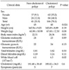
Of the 187 patients, 79 patients were female and 108 were male. The percentage of cholesterol polyps were higher in both sexes (78.48% in female, 77.77% in male) but there were no statistically significant differences in the gender ratio between the cholesterol polyp group (M:F = 1.35:1) and the non-cholesterol polyp group (M:F = 1.41:1). The mean ages were 41.59 ± 11.36 for the cholesterol group and 46.37 ± 12.89 for the non-cholesterol group, which showed significantly different results between the two groups (P = 0.024) (Table 1).
Twelve patients had presenting symptoms of right upper quadrant pain and discomfort (7 in cholesterol polyps, 5 in non-cholesterol polyps). One hundred and fifty-seven cases were associated with chronic cholecystitis (130 in cholesterol polyps, 27 in non-cholesterol polyps). Forty-three cases of cholesterol polyps (29%) and 9 cases for non-cholesterol polyps were associated with a GB stone. The mean BMI was 24.70 ± 3.18 in the cholesterol polyp group and 22.70 ± 2.91 in the non-cholesterol polyp group, which was significantly different between the two groups (P = 0.01) [13,14]. The cholesterol level was 185.02 ± 34.3 in the cholesterol polyp group and 181.48 ± 38.45 in the non-cholesterol polyp group but there were no significant differences between the two groups (P=0.6) (Table 1).
The data related with operation in both cholesterol polyp and non-cholesterol polyp were evaluated and showed no significant difference between the two groups (operation time, hospital day, postoperative day, conversion rate). Four cases of the cholesterol polyp group and 1 case of the non-cholesterol polyp group were considered to be open on the preoperative state because of the history of previous abdominal surgery and the consideration of ductal damage due to adhesion (Table 2).
Of the 187 cases, 146 (78.1%) were cholesterol polyps [11] and 41 (21.9%) were non-cholesterol polyps. Of the 41 non-cholesterol polyps 2 were inflammatory polyps and 2 were fibrous polyps, 9 hyperplastic polyps, 27 adenomatous polyps (65.85%) and 1 malignant polyp (Table 3) [10].
As a result, 59.59% of the cholesterol polyps was multiple. In contrast, 90.24% was solitary polyps in the non-cholesterol polyp group. But the percentage of associating GB stones were almost the same between the two groups (29% in cholesterol polyp group, 22% in non-cholesterol polyp group) (Table 4).
The preoperative mean maximum diameters measured by the USG in the cholesterol group and the non-cholesterol group were 10 ± 3.2 mm and 10 ± 4.28 mm, retrospectively; whereas by CT they were 9 ± 4.73 mm and 9 ± 5.56 mm, retrospectively. The mean diameters from CT scanning tended to be smaller than from the USG in both groups.
The mean maximum diameters measured pathologically were 5 ± 3.00 mm in cholesterol polyps and 10 ± 4.37 mm in non-cholesterol polyps. The correlation coefficient between USG, CT, and pathologic size was compared in both groups shown to be 0.71 cm (P = 0.000) in the non-cholesterol polyp group and 0.082 cm (P = 0.167) in the cholesterol polyp group.
Of the non-cholesterol polyp group the correlation coefficient between the USG and the CT was 0.708 (P = 0.000) while it was 0.422 (P = 0.001) in the cholesterol polyp group. The correlation coefficient between the USG and pathology was 0.625 (P = 0.000) in the non-cholesterol polyp group and 0.331 (P = 0.001) in the cholesterol polyp group (Tables 5, 6).
Unfortunately, 28.6% of the cholesterol polyps was not found in the preoperative CT, which was able to be detected on the USG. But the percentage of the undetectable rate was significantly lower (8%, P = 0.032) than in the non-cholesterol polyp group. This means, if the preoperative radiologic finding shows polyps only in the USG, it can be more likely a cholesterol polyp than a non-cholesterol polyp (Table 7).
Though a laparoscopic cholecystectomy can be considered a less complicated operation, an operation that is unnecessary is truly a burden to the patient and a waste of cost. To prevent these operations, the diagnosis of GB polyps has to be made correctly. This study was intended to characterize the clinical features of the cholesterol polyp and to determine the accurate radiological predictive factors.
Age is a known risk factor associated with malignancy [13-15]. This study also showed a higher mean age in the non-cholesterol polyp group. Many studies show the relationship between metabolic syndrome and the development of cholesterol polyps [2,16,17]. Our study showed a significant data of higher BMI in cholesterol polyps compared to non-cholesterol polyps [11]. And the cholesterol level was higher in the cholesterol polyp group without significant difference.
It is known that a single polyp is more likely to be a malignant polyp [13,18]. If a single polyp is identified it needs more aggressive work up or more aggressive interventions than multiple polyps. Among our study populations, over 90% of the non-cholesterol polyps was solitary and about 60% of cholesterol polyps was multiple polyps. In other words our study also shows a need for aggressive work up in solitary polyps as the ratio is higher in non-cholesterol polyps. Many studies have reported that polyps over 10 mm have a high risk of being a malignancy, and this is surgical criteria for treating GB polyps [11,19-21]. The pathologic size of the polyps in our study also had a similar tendency. But there were many cholesterol polyps larger than 10 mm in the group. Therefore, it is hard to state size as a definite factor to distinguish benign from malignant polyps [22-25].
We observed discrepancies between preoperative radiological measurements and postoperative pathologic measurements in the cholesterol polyp group. And the cholesterol polyp group also showed low correlation coefficients between the three measured sizes. The damage of cholesterol polyps during operation or handling after obtaining the GB may explain the results. As a result, the preoperative radiologic studies are limited in obtaining the correct measurements for cholesterol polyps. On the contrary, non-cholesterol polyps have a high correlation coefficient between the preoperative diameters measured by USG and CT. And the correlation coefficiency compared with the USG and CT of postoperative pathologic diameters are also significantly high.
The predictive preoperative radiologic signs for a non-cholesterol polyp include a single polyp, observable on both the USG and CT, and a discrepancy of the maximum diameter less than 1 mm between the USG and CT.
In conclusion, young patients with high BMI and high cholesterol levels who have multiple GB polyps near 10 mm in size measured by USG can be recommended a CT scan [26-28]. And if the diameter has a discrepancy or only seen in the USG, the GB polyp can be considered as a cholesterol polyp and require planning for another follow-up after 6 months [29,30] thereby delaying the operation. We suggest that it would be more efficient to make flexible treatment plans for the mentioned cases rather than the fixed guideline.
References
1. Chen CY, Lu CL, Chang FY, Lee SD. Risk factors for gallbladder polyps in the Chinese population. Am J Gastroenterol. 1997. 92:2066–2068.
2. Segawa K, Arisawa T, Niwa Y, Suzuki T, Tsukamoto Y, Goto H, et al. Prevalence of gallbladder polyps among apparently healthy Japanese: ultrasonographic study. Am J Gastroenterol. 1992. 87:630–633.
3. Onoyama H, Yamamoto M, Takada M, Urakawa T, Ajiki T, Yamada I, et al. Diagnostic imaging of early gallbladder cancer: retrospective study of 53 cases. World J Surg. 1999. 23:708–712.
4. Lee KF, Wong J, Li JC, Lai PB. Polypoid lesions of the gallbladder. Am J Surg. 2004. 188:186–190.
5. Jorgensen T. Gall stones in a Danish population. Relation to weight, physical activity, smoking, coffee consumption, and diabetes mellitus. Gut. 1989. 30:528–534.
6. Barbara L, Sama C, Morselli Labate AM, Taroni F, Rusticali AG, Festi D, et al. A population study on the prevalence of gallstone disease: the Sirmione Study. Hepatology. 1987. 7:913–917.
7. Sadamoto Y, Oda S, Tanaka M, Harada N, Kubo H, Eguchi T, et al. A useful approach to the differential diagnosis of small polypoid lesions of the gallbladder, utilizing an endoscopic ultrasound scoring system. Endoscopy. 2002. 34:959–965.
8. Azuma T, Yoshikawa T, Araida T, Takasaki K. Differential diagnosis of polypoid lesions of the gallbladder by endoscopic ultrasonography. Am J Surg. 2001. 181:65–70.
9. Choi WB, Lee SK, Kim MH, Seo DW, Kim HJ, Kim DI, et al. A new strategy to predict the neoplastic polyps of the gallbladder based on a scoring system using EUS. Gastrointest Endosc. 2000. 52:372–379.
10. Sugiyama M, Xie XY, Atomi Y, Saito M. Differential diagnosis of small polypoid lesions of the gallbladder: the value of endoscopic ultrasonography. Ann Surg. 1999. 229:498–504.
11. Koga A, Watanabe K, Fukuyama T, Takiguchi S, Nakayama F. Diagnosis and operative indications for polypoid lesions of the gallbladder. Arch Surg. 1988. 123:26–29.
12. Shinkai H, Kimura W, Muto T. Surgical indications for small polypoid lesions of the gallbladder. Am J Surg. 1998. 175:114–117.
13. Yeh CN, Jan YY, Chao TC, Chen MF. Laparoscopic cholecystectomy for polypoid lesions of the gallbladder: a clinicopathologic study. Surg Laparosc Endosc Percutan Tech. 2001. 11:176–181.
14. Terzi C, Sokmen S, Seckin S, Albayrak L, Ugurlu M. Polypoid lesions of the gallbladder: report of 100 cases with special reference to operative indications. Surgery. 2000. 127:622–627.
15. Jorgensen T, Jensen KH. Polyps in the gallbladder: a prevalence study. Scand J Gastroenterol. 1990. 25:281–286.
16. Sahlin S, Granström L, Gustafsson U, Stählberg D, Backman L, Einarsson K. Hepatic esterification rate of cholesterol and biliary lipids in human obesity. J Lipid Res. 1994. 35:484–490.
17. Sandri L, Colecchia A, Larocca A, Vestito A, Capodicasa S, Azzaroli F, et al. Gallbladder cholesterol polyps and cholesterolosis. Minerva Gastroenterol Dietol. 2003. 49:217–224.
18. Doh YW, Lee JH, Lim HM, Chi KC, Park YG. Polypoid lesions of gallbladder: clinicopathological features and indication of operation. J Korean Surg Soc. 2005. 69:245–251.
19. Akatsu T, Aiura K, Shimazu M, Ueda M, Wakabayashi G, Tanabe M, et al. Can endoscopic ultrasonography differentiate nonneoplastic from neoplastic gallbladder polyps? Dig Dis Sci. 2006. 51:416–421.
20. Mainprize KS, Gould SW, Gilbert JM. Surgical management of polypoid lesions of the gallbladder. Br J Surg. 2000. 87:414–417.
21. Yang HL, Sun YG, Wang Z. Polypoid lesions of the gallbladder: diagnosis and indications for surgery. Br J Surg. 1992. 79:227–229.
22. Pandey M, Sood BP, Shukla RC, Aryya NC, Singh S, Shukla VK. Carcinoma of the gallbladder: role of sonography in diagnosis and staging. J Clin Ultrasound. 2000. 28:227–232.
23. Levy AD, Murakata LA, Abbott RM, Rohrmann CA Jr. From the archives of the AFIP. Benign tumors and tumorlike lesions of the gallbladder and extrahepatic bile ducts: radiologic-pathologic correlation: Armed Forces Institute of Pathology. Radiographics. 2002. 22:387–413.
24. Csendes A, Burgos AM, Csendes P, Smok G, Rojas J. Late follow-up of polypoid lesions of the gallbladder smaller than 10 mm. Ann Surg. 2001. 234:657–660.
25. Sugiyama M, Atomi Y, Yamato T. Endoscopic ultrasonography for differential diagnosis of polypoid gall bladder lesions: analysis in surgical and follow up series. Gut. 2000. 46:250–254.
26. Kim SJ, Lee JM, Lee JY, Choi JY, Kim SH, Han JK, et al. Accuracy of preoperative T-staging of gallbladder carcinoma using MDCT. AJR Am J Roentgenol. 2008. 190:74–80.
27. Sadamoto Y, Kubo H, Harada N, Tanaka M, Eguchi T, Nawata H. Preoperative diagnosis and staging of gallbladder carcinoma by EUS. Gastrointest Endosc. 2003. 58:536–541.
28. Weaver SR, Blackshaw GR, Lewis WG, Edwards P, Roberts SA, Thomas GV, et al. Comparison of special interest computed tomography, endosonography and histopathological stage of oesophageal cancer. Clin Radiol. 2004. 59:499–504.
29. Boulton RA, Adams DH. Gallbladder polyps: when to wait and when to act. Lancet. 1997. 349:817.
30. Ilias EJ. Gallbladder polyps: how should they be treated and when? Rev Assoc Med Bras. 2010. 56:258–259.


 ePub
ePub Citation
 Print
Print


 XML Download