

 ePub
ePub Citation
 Print
Print


INTRODUCTION
Acute appendicitis is the most common disease requiring emergency surgery in children. In numerous studies, when conventional laparoscopic appendectomy (C-LA) using 3 ports is compared with open appendectomy, it has advantages of reduced pain, reduced hospital stay, and enhanced cosmetic effects [1-3]. Recently, as technology and innovation continue to advance the field of minimally invasive surgery, single incision laparoscopic surgery (SILS) is being applied to diverse surgeries as a new technique for minimal invasive surgery [4-7].
In studies comparing single incision laparoscopic surgery for appendectomy (SILS-A) with a C-LA in adults, although early pain was observed, the former was superior from a cosmetic viewpoint, and the incidence of complications was not different. Thus, recently, it was reported as a technique that could be performed safely in adults [8-10].
However, studies on the application of SILS-A in children are few, and recently, Oltmann et al. [11] showed that SILS-A operating times in patients with non-perforated appendicitis are somewhat longer than with C-LA, but should decrease with improved instrumentation and experience.
Therefore, we performed this study to examine the feasibility and efficacy of SILS-A in children by comparing SILS-A with C-LA.
METHODS
The study was performed on 55 cases of appendicitis in adolescent patients who underwent either a conventional 3-port laparoscopic appendectomy or single incision laparoscopic surgery for appendectomy (SILS-A) by the same surgeon from July 2009 to March 2011 at our hospital. Patients receiving a C-LA were 25 cases, and SILS-A were 30 cases.
Among the patients who underwent SILS-A were 8 cases of complicated appendicitis due to perforation or gangrenous appendicitis; 5 cases of which had one additional port inserted to facilitate the manipulation of laparoscopy or drainage.
This study was a retrospective review of medical records. The technique of SILS-A was approved by the Ethical Committee of our hospital.
Prior to surgery, abdominal ultrasonography or computed tomography was performed on all patients. In regard to surgical methods, C-LA, SILS-A, and laparotomy, were explained to the guardians, after which the method was selected by the patients themselves and their guardians.
Complicated appendicitis is defined as cases showing gangrene or perforation changes detected by surgical findings or histological findings, or cases with an abscess in the vicinity of the appendix.
General anesthesia was administered to all patients. Simultaneously with the diagnosis of appendicitis, an antibiotic, 2nd generation cephalosporin, was administered; and for cases diagnosed as appendicitis associated with complications, aminoglycoside and metronidazole were also administered.
In the supine position, the surgeon stood at the left lower area of the patient, leaning toward the lower extremities, and the first assistant manipulated the laparoscope on the right upper side of the surgeon. C-LA was performed using 3-trocar techniques, a 10-mm trocar was inserted through the vicinity of the umbilicus, a 5-mm trocar was inserted between the pubic bone and the middle of the umbilicus, and another 5-mm trocar was inserted in the vicinity of the McBurney point. The mesoappendix was ligated and dissected by the application of a LigaSure (Valleylab, Boulder, CO, USA) and electric coagulation. The appendiceal base was ligated by the use of one Endo loop (Ethicon Inc., Somerville, NJ, USA).
To prevent infection in the area of the trocar insertion, the surgeon removed the resected appendix to the extracorporeal area using a Lap-bag (Sejong Medical, Paju, Korea) through the 10-mm trocar area. The abdominal cavity was washed with saline. Afterward, for cases showing perforation or severe inflammation, such as an abscess in the vicinity of the appendix, sufficient drainage after surgery was achieved by installing a Jackson-pratt drain through the 3rd trocar.
SILS-A was performed in the supine position under general anesthesia, and in the umbilical area, according to the open incision method, a 1.5- to 2-cm vertical incision was made. If the umbilical area was severely dirty or malodorous, avoiding the center of the umbilical area, in the area above the umbilical area or based on the umbilical area, a half-moon incision window 1.5- to 2-cm in size was made. When the insertion route to the abdominal cavity was secured, a wound retractor (Alexis, Applied Medical Resources Co., Rancho Santa Margarita, CA, USA) was inserted. One 5-mm trocar for use with a 30°, 5-mm laparoscopic camera (Karl-Storz, Tuttlingen, Germany) and the injection of CO2 gas, two homemade 5-mm trocars to reduce collisions of the tips of the trocars during SILS-A, and a three-way catheter to remove smoke generated during the use of the electric coagulator were fixed using silk in the finger area of the surgical gloves to prevent the leakage of the air.
For cases difficult to resect because of perforation or severe inflammation, such as an abscess in the vicinity of the appendix and requiring drainage, an additional 5-mm trocar was inserted in the vicinity of the McBurney point.
Meso-appendectomy and appendectomy were performed by using identical conventional laparoscopic methods or extra-corporeal appendectomy when the cecal base can be mobilized to the midline.
In the 6 cases of intra-corporeal SILS-A, after the resection of the appendix, the resected appendix was added to the finger of the glove that was no longer required and ligated with a forcep.
If the appendix was big or contamination was severe, it was removed to the extracorporeal area using the Lap-bag; the abdominal cavity was washed, and the wound retractor was removed. If drainage was required, a 5-mm trocar was inserted in the vicinity of the McBurney point, and a Jackson-pratt drain was installed.
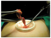
In the 24 cases of extra-corporeal SILS-A, the tip of the appendix or mesentery is grasped. The insufflation is released and the appendix is extruded through the umbilicus while removing the glove. The appendix is brought out of the wound until the cecal base can be grasped with a Babcock clamp and appendectomy is performed in the standard open fashion (Fig. 1).
In all patients, a patient-controlled analgesia (Accufusor, WooYoung Medical, Jincheon, Korea) was used. The patient-controlled analgesia, 18 µg/kg of fentanyl and 3 mg/kg of Keromin (Ketorolac Tromethamine, Hana Pharm Co., Hwasung, Korea) were diluted with metoclopramide and saline to a 100-mL volume and injected. For cases presenting with severe pain, higher than 5 points on a verbal numerical rating scale, despite the use of patient-controlled analgesia, as additional analgesic, Keromin was injected intravenously.
Statistical analysis was performed by using the Student's t-test and the chi-square test with the SPSS ver. 17.0 (SPSS Inc., Chicago, IL, USA). P-values lower than 0.05 were considered to be statistically significant.
RESULTS
The ratio of males to females for the patients who underwent SILS-A was 17 : 13; their mean age was 9.3 ± 4.0 years. In the group that underwent a C-LA, the ratio of males to females was 14 : 11; their mean age was 8.7 ± 3.5 years.
In patients who underwent SILS-A, 8 patients had complicated appendicitis; additional trocar had to be inserted for severe inflammation, abscess and drainage (5 patients), but none of those cases were converted to 3-port laparoscopic appendectomy or a laparotomy. In the group that underwent a C-LA, 7 patients had complicated appendicitis; none of those cases were converted to laparotomy.
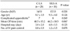
The operation time of the group that underwent SILS-A was 46.2 ± 18.5 minutes; C-LA was 40.5 ± 15.2 minutes. Although the time was longer for the group that underwent SILS-A, no statistically significant differences were detected (P = 0.067).
The hospitalization periods after surgery of the group that underwent SILS-A were 4.0 ± 1.5 days, and that of the group that underwent C-LA, 3.8 ± 2.0 days. The hospitalization period showed no statistically significant difference.
The frequency of additional analgesics administered to SILS-A group was 1.2 ± 1.5 times, and that for C-LA group was 0.8 ± 0.5 times. The frequency of additional analgesics in SILS-A group was higher than C-LA but showed no statistically significant difference (P = 0.078) (Table 1).
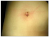
In regard to post-operative complications, in SILS-A group, seroma in the umbilical area developed in 2 patients, and in C-LA group, seroma and ileus developed simultaneously in 1 patient. They recovered after conservative management (Table 2). Fig. 2 is immediate postoperative scar after SILS-A in a 9-year-old female patient with gangrenous type appendicitis.
DISCUSSION
Since the first laparoscopic appendectomy was reported by Semm [12] in Germany for an appendix without inflammation, it has been performed by numerous surgeons. In comparison with open appendectomy, laparoscopic appendectomy have the benefits of reduction of postsurgical pain, decreased operative trauma resulting in quicker recovery, shorter hospital stays, and improved cosmesis. As a result, it is now widely performed in adults as well as pediatric patients by many practicing surgeons [1-3].
As laparoscopic minimal invasive surgery draws attention, interest in no-scar surgical methods is on the rise. Together with the development of equipment, Natural Orifice Transluminal Endoscopic Surgery, single-trocar or single incision surgical methods have been applied to diverse diseases in the abdominal cavity [4-7,13]. Although it differs slightly depending on the surgeon, single incision laparoscopic surgery for appendectomy makes an incision window through the umbilicus in most cases. It is applied to appendectomy as a new technique of minimal invasive surgery because the umbilicus is located in the middle of the abdomen, so diverse intra-abdominal approaches can be performed; blood vessels and nerves are absent, so incision windows can be readily created; even after surgery, wounds become depressed within the umbilicus and, thus, may be considered as an existing congenital scar [8-10,14].
Reviewing the reports that compared single incision laparoscopic surgery with a conventional 3-port laparoscopic appendectomy in adults, the former was found to reduce scars in addition to having the advantages of a 3-port laparoscopic appendectomy; thus, it is advantageous for cosmetic improvement. Nonetheless, shortcomings, long operation time, and substantial early post-surgical pain, have been reported [8-10].
Oltmann et al. [11] reported that single incision laparoscopic surgery for appendectomy is both feasible and safe across the pediatric age range. Although operating room times are somewhat longer than with conventional 3-port laparoscopic appendectomy, they concluded that it should decrease with improved instrumentation and experience.
To overcome the longer operative time, we used a 30°, 5-mm laparoscopic camera to minimalize collisions with and interference between the laparoscopic surgical equipment and the laparoscopic camera. For cases in which collision and interference phenomenon between laparoscopic surgical equipment and laparoscopic cameras occur, in the view of 30° - 5-mm laparoscopic cameras, laparoscopic manipulation was made easy by using the flexible laparoscopic Roticulator Grasper, Dissector, and Shear (Covidien, Norwalk, CT, USA).
Finally, we adapted extra-corporeal appendectomy when the cecal base could be mobilized to the midline. In 24 out of 30 patients (80%) extra-corporal appendectomy was applicable, but in 6 patients (20%) it was not applicable due to non-mobile cecum and adhesion. We think that it is an important point to choose intra or extra-corporeal appendectomy whether the cecum is mobile or not. In the cases of mobile cecum, extra-corporeal appendectomy method in adolescent patients is a appropriate method to avoid unnecessary manipulation and reduce operation time.
Extra-corporeal laparoscopic appendectomy using single umbilical incision, initially published by Pelosi and Pelosi [15], may offer some advantage in terms of expense. Removal of the appendix extracorporeally in the manner of conventional surgery eliminates need for expensive devices. Visnjic [16] reported that transumbilical extra-corporeal laparoscopically assisted appendectomy operative time in children was shorter and cost less than conventional 3 port laparoscopic appendectomy. Hence, they called this method "High-tech low-budget surgery."
Through such methods as those discussed above, single incision laparoscopic surgery for appendectomy in children can even be applied to appendicitis patients; and the operation time may not be significantly longer.
Kang et al. [9] reported that early pain was more severe in single incision laparoscopic surgery for appendectomy in adults than it was in a conventional 3-port laparoscopic appendectomy. This might be caused by the fact that although the skin incision in the umbilical area is small, the actual length of the fascia incision is longer, and through a small incision window, laparoscopic equipment is used simultaneously, which irritates the incision window.
Visnjic [16] also reported that in transumbilical extra-corporeal laparoscopically assisted appendectomy in children, the administration of rescue analgesia was not statistically different than in conventional 3-port laparoscopic appendectomy.
In our study, similarly, in the single incision laparoscopic surgery for appendectomy in children, analgesic administration was significantly greater, and this is thought to be associated with shorter operation time and less fascial irritation by performing extra-corporeal laparoscopic appendectomy.
Postoperative complications in patients who underwent single incision laparoscopic surgery for appendectomy were treated without significant side effects or complications, except wound problems. A seroma in the umbilical area developed in 2 patients (6.6%) and treated in outpatient clinics.
In the report of Oltmann et al. [11], they reported the incidence of wound complication was 5.2% (1/19), and Visnjic [16] was 13.7% (4/29). The higher incidence of port site infection could be expected in single incision laparoscopic surgery for appendectomy than conventional 3-port laparoscopic appendectomy, especially in extracorporeal appendectomy due to the exposure and manipulation of the appendix on the incision site. Therefore, an adjusted operative technique using minimal, gentle movements, and adequate wound protection is required. We routinely used a wound retractor for wound protection, and wound seroma, not wound infection, developed in only 2 cases.
In conclusion, single incision laparoscopic surgery for appendectomy in children is technically feasible and safe. Considering little postoperative scar and no difference of post-operative outcomes compared to conventional 3-port laparoscopic appendectomy, single incision laparoscopic surgery for appendectomy in children could be applicable. Larger studies and further technical implements will be necessary to assess the true benefit of this approach.

 XML Download