

 Citation
 Print
Print


INTRODUCTION
Paraffinoma is a chronic granuloma, caused by the prosthetic or therapeutic injection of paraffin into tissues. It usually presents as a firm, discrete mass that can be extremely painful and disfiguring. Other complications of paraffinoma include edema, induration, ulceration, and skin necrosis. Paraffinoma may occasionally mimic neoplasm on physical examination and imaging studies [1], and may complicate ultrasonographic examination with posterior shadowing artifacts [2].
We describe a case of difficult preoperative ultrasonographic evaluation of the thyroid gland due to previous cervical paraffin injection. Written informed consent was obtained from the patient for publication of this case report.
CASE REPORT
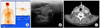
A 60-year-old woman was presented with a thyroid nodule that was incidentally discovered on routine medical check-up. The patient underwent a fluorine-18-fluorodeoxyglucose positron emission tomography scan at a local hospital and abnormal hot uptake was discovered in the left thyroid gland (Fig. 1A). She had no specific relevant medical history, except for paraffin injection into the neck for a neck lift 10 years ago.
On physical examination, multiple small nodules were noted in the neck. Cervical ultrasonography showed multiple granulomas in the neck and around the thyroid gland that limited evaluation of the gland. Because of the limited view, ultrasonography-guided fine needle aspiration cytology failed to diagnose thyroid carcinoma. Computed tomography (CT) demonstrated solid and cystic masses in the isthmus and left lobe of the thyroid gland that appeared suspicious for cancer (Fig. 1B, C).
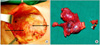
A diagnostic left hemithyroidectomy was performed for suspicion of thyroid carcinoma. Operatively, two nodules were found in the isthmus and left thyroid measuring 1.3 cm and 1.5 cm in diameter, respectively. Using an intraoperative frozen section, papillary thyroid carcinoma was confirmed in the left thyroid mass and total thyroidectomy was performed immediately. In addition, two large palpable granulomas in the neck were removed (Fig. 2).
Postoperative histopathological examination confirmed classic papillary thyroid carcinoma in the left thyroid and papillary thyroid carcinoma with nodular fasciitislike stroma in the isthmus.
The patient was discharged from the hospital four days after surgery without any complications. She underwent postoperative low-dose radioactive iodine treatment for ablation, and an iodine-131 whole body scan showed no abnormal uptake in the thyroid bed or other areas.
The patient has received routine check-ups and measurements of serum thyroglobulin concentrations along with thyroid stimulating hormone suppression therapy. Follow up ultrasonography was performed at 6 months after the operation, and there was no difficulty in evaluation the thyroid operation bed.
DISCUSSION
Attempts to alter the body contour have been made since the 19th century, with numerous foreign materials introduced into different areas of the body [3]. Subcutaneous injections of high viscosity fluids have been used for more than a century to augment and expand tissues. Paraffin, a colorless hydrocarbon mixture commonly used to make candles and lubricants, was first introduced in 1890 as a cheap and quick method of breast augmentation.
Paraffinoma is a granulomatous resection resulting from the injection of a foreign substance containing straight-chain saturated hydrocarbons such as paraffin or mineral oil. The body lacks the enzymes to metabolize interstitial exogenous oils, and a foreign body reaction occurs [4].
Macroscopically, excised specimens are white or grayish-white. Microscopic changes manifest macroscopically in the form of undesired reactions including inflammation, edema, scarring, necrosis, deformity, ulceration, and sterile abscesses [3,5]. There is granulomatous inflammation with multiple clear vacuoles, resulting in a "Swiss cheese" appearance [6]. The histopathological features include thickening of the reticular dermis and replacement of normal subcutaneous fat by lakes of oil interspersed with fibrous tissue and a granulomatous chronic inflammatory reaction [5].
Although the histopathological examination was not confirmed in our patient, the diagnosis of paraffinoma made sense in light of the physical examination findings and medical history.
Ultrasonography is the imaging modality of choice for preoperative evaluation and characterization of thyroid lesions in thyroid cancer [7]. Fine-needle aspiration cytology is the next step in the diagnosis of thyroid cancer, and is usually performed under ultrasonographic guidance for exact targeting of the lesions.
However, paraffinomas, which exhibit marked posterior acoustic shadowing, can complicate ultrasonographic examination of the thyroid gland and therefore limits the usefulness of ultrasonography in investigating thyroid tumors in patients with neck paraffinomas [2].
In the present case, ultrasonographic evaluation was complicated by posterior acoustic shadowing of the implants and we chose to perform neck CT. The results of the CT scan were not diagnostic but were helpful for localization of the lesion in the thyroid gland when taken along with other pertinent information [8]. We performed ipsilateral lobectomy and an intraoperative frozen section and finally confirmed the diagnosis of papillary thyroid carcinoma.
Independent of the paraffinoma, a rare variant of nodular fasciitis-like stroma was confirmed postoperatively. Only 18 cases of this variant have been reported in the English literature [9]. The paraffin injection was not histologically correlated with this rare variant.
In conclusion, radiologic findings in the patients with previous paraffin injections may be compromised by artifacts and lead to diagnostic confusion or misdiagnosis. Awareness of this form of augmentation and obtaining a pertinent patient history will help clinicians establish the correct diagnosis. If needed, diagnostic modalities including CT, magnetic resonance imaging, and intraoperative frozen section should be performed to confirm the diagnosis.
 XML Download