

 Citation
 Print
Print


INTRODUCTION
Although branchial cleft cysts (BCCs) are common, squamous cell carcinomas of BCC are rare [1], but papillary carcinomas arising from them are extremely rare. Here we report a patient with papillary carcinoma originated from right lateral BCC without any evidence of a papillary carcinoma in thyroid gland. Papillary carcinoma of BCC may be clinically indistinguishable from benign BCC or metastatic papillary carcinoma, and the diagnosis in most cases is incidental after surgical resection. We report the 10th case for papillary carcinoma in a BCC, and review the literature on the features of the disease and discuss the role of immunohistochemical staining with thyroglobulin (TG), thyroid-associated transcription factor-1 (TTF-1) and p63.
CASE REPORT
A 41-year-old woman presented to our hospital with a 2-year history of palpable mass in the right lateral aspect of the neck. There was no past history of irradiation or trauma in the head and neck. On physical examination, a firm and non-tender mass was palpable in the right lateral neck measuring 3.0 cm at the level of the cricoid cartilage without associated cervical lymphadenopathy. No abnormal laboratory findings were seen in routine laboratory investigations and euthyroid state in thyroid function test.
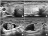
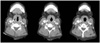
Neck ultrasonography revealed a solitary nodule measuring 0.5 cm in the right thyroid gland, and a 3.0 cm mixed solid and cystic mass in the right lateral aspect of the neck at level III (Fig. 1). Computed tomography also showed mixed solid and cystic lesion associated with punctate calcification (Fig. 2).
Examination of fine needle aspiration cytology (FNA) on thyroid gland confirmed follicular cell proliferative lesion. But, the right lateral neck mass was suggestive of metastatic papillary carcinoma. Therefore, the patient was diagnosed with presumed metastatic papillary thyroid carcinoma (PTC) in the right lateral neck and occult papillary thyroid carcinoma in the right thyroid gland.
The patient underwent right lateral neck dissection followed by total thyroidectomy. It must be emphasized that papillary carcinoma arising in a right lateral BCC was not in the differential diagnoses preoperatively. The intraoperative findings included a 3.0 cm dark brown fluid filled cystic mass lateral to the internal jugular vein without cyst rupture at the time of the operation, and revealed PTC on the frozen section biopsy. Therefore, a total thyroidectomy was followed.
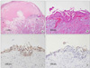
Histopathologic examination of the 3.0 cm cystic mass was confirmed papillary carcinoma in a BCC (Fig. 3A). The cyst was partially lined by squamous epithelium surrounded by fibrous tissues associated with chronic inflammation (Fig. 3B). But there was no normal thyroid tissue adjacent to the focus of papillary carcinoma within the cyst wall (Fig. 3A).
Histopathologic examination of the thyroid gland revealed nodular hyperplasia of the right lobe without evidence of malignancy. Repeated serial sectioning of the whole thyroid and even in postoperative positron emission tomography does not reveal any foci of carcinoma. Other harvested right lateral lymph nodes were examined but there were no evidence of any other abnormal findings.
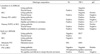
It is known that immunohistochemical staining through TG, TTF-1 and p63 could help in the differential diagnosis in thyroglossal duct cyst, BCC, and papillary carcinoma in BCC (Table 1). In our case, the squamous epithelium was positive for p63 and negative for TG and TTF-1 (Fig. 3C, D). On the other hand, papillary carcinoma in BCC was positive on TG and TTF-1, but negative on p63. This issue for immunohistochemical staining will be explained later on discussion.
The patient has remained disease free having been followed up for 28 months. She is being managed with post-operative levothyroxine replacement without radioactive iodine therapy.
DISCUSSION
Ectopic thyroids are rarely found in the region of the sublingual, submandibular, intratracheal, mediastinum, esophagus, lung, heart, aorta, and even in abdomen. The most frequent site is the thyroglossal remnants or BCC. In our knowledge, about 215 cases papillary carcinoma of thyroglossal remnants were reported, but only nine cases of BCC have been reported by 2010 [2-4].
Because metastatic papillary carcinoma may become cystic [5], all lining cells of the cyst must be carefully reviewed. And it is important to differentiate a metastatic derivation with a missed primary tumor or represents papillary thyroid carcinoma arising in ectopic thyroid tissue in a BCC [5-7].
Sidhu's criteria for the papillary carcinoma in BCC [3] was suggested as 1) an epithelial lining layer, subepithelial lymphoid tissue collection, 2) normal thyroid tissue adjacent to the focus of papillary carcinoma within the wall, 3) and no evidence of papillary carcinoma in the thyroid or other area.
In our case, there was no normal thyroid tissue adjacent to the papillary carcinoma within the wall. It may be assumed as 1) papillary carcinoma was initiated in the normal thyroid tissues in BCC, 2) and all normal thyroid tissues within the wall were replaced by papillary carcinoma. But there was epithelial lining layer - not in cystic change of metastatic carcinoma -, and there was no evidence of papillary carcinoma within the thyroid gland even in the repeated serial section and immunohistochemical staining.
In immunohistochemical staining, TTF-1 cannot distinguish between primary and metastatic tumors of BCC [8]. The p63 is a potentially oncogenic protein that would contribute to the onset of papillary carcinoma. It was suggested that he detection of p63 in papillary carcinomas of BCC could distinguish from primary BCC or metastatic origin [6]. Unfortunately, p63 staining on papillary area of carcinoma of BCC in our case did not display any positive foci. As reviewed by Lanzafame et al [6], the involvement of p63 immunoreactive cells in the tumorigenic process remains to be proven because they may simply reflect squamous differentiation of papillary thyroid carcinoma. In addition, staining of p63 was focal rather than extensive or confluent, and the extent of staining varied from multiple foci to scattered or rare foci. It was also in only one papillary type carcinoma in BCC case, and follicular variant carcinoma of BCC in his study showed negativity on p63 [6]. We need some more research for this debate on positivity on p63 in PTC of BCC.
In our case, there was no evidence of papillary carcinoma found within the thyroid gland when meticulous histological examination including immunohistochemical staining on all thyroid section. Also, diagnostic radioactive iodine scan and positron emission tomography does not reveal the any focus of malignancy. Taken together, these findings confirmed papillary carcinoma arising from the BCC at level III of the right neck even though absence of normal thyroid tissue within the BCC.
In heterotopic thyroid tissue in thyroglossal duct cyst and BCC, it is important to evaluate if the case has a metastatic of occult thyroid cancer. So FNA should be performed under ultrasound guidance in order to best sample the solid part of the lesion and any thyroid nodule in any size. This can help to reduce false-negative rate from dilution by the cystic contents. and TG levels in FNA fluids can aids the adequate diagnosis and decision of operation extent [4]. Papillary carcinoma arising from branchial remnants has the ability to metastasize to regional lymph nodes. Neck node metastases are found in 20% [9,10], but distant metastases were not reported.
In conclusion, it should be emphasized that the surgeon must be cautioned that the possibility of primary papillary carcinoma in the branchial cleft cyst.
 XML Download