

 ePub
ePub Citation
 Print
Print


INTRODUCTION
Gastrointestinal melanosis is discovered incidentally during endoscopic studies, and occurs most in the colon. In rare cases, it can also develop in the ileum, duodenum or esophagus. From among these, melanosis ilei does refer to the deposition of grayish-black or brownish-black color pigments in the ileal mucosa grossly. In the past, causative materials of melanosis were speculated to be the melanin. However, with the use of electron microscopy, electron probe energy dispersive X-ray analysis studies and related techniques, it has been revealed that the causative materials are different based on the affected gastrointestinal tract organ [1-3].
Worldwide, the reports on melanosis ilei are very rare. Since Yoon et al. [4] report a case of melanosis ilei by charcoal ingestion, only 3 cases by charcoal ingestion and 1 case by oral iron had been reported in Korea until now [4-7].
Few researchers have investigated whether the black pigment lesion of melanosis ilei are continuous or discontinuous, whether they tend to occur in the entire small intestine or only in the terminal ileum and whether the pigments disappear if the exposure to causative materials is discontinued. Below we present the case in detail, together with a review of the relevant literature.
CASE REPORT
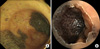
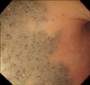
A 65-year-old female patient visited our hospital for chronic diarrhea, which developed 3 years prior to the appointment, as the chief complaint. For the treatment of intermittent abdominal pain, dyspepsia and diarrhea, the patient had ingested 10 g/day commercial edible charcoal over a three-year period of time. Her past medical history was not significant for any special finding, nor was her social or family histories. The patient was employed as a housekeeper at the time of evaluation. When queried about drug history, the patient reported no history of smoking, laxatives, or ingestion of herbal medicines. At the time of visit, blood pressure was 102/80 mmHg, pulse rate was 70 times/min, respiration rate was 20 times/min and body temperature was 36.3℃. During palpation, mild tenderness in the lower abdomen was detected. In laboratory findings, blood cell counts and blood chemistry showed normal findings. During the colonoscopy, the large intestine showed normal findings, and in the terminal ileum, several black colored geographic mucosal pigmentations were observed. The boundaries of the lesions were well demarcated, and the adjacent mucosa showed normal characteristics. During the gastroduodenoscopy, mucosal pigmentation was not observed in either the esophagus or the duodenum. To examine the small intestine whether this lesion involved entire small intestine or not, capsule endoscopy was performed, and black pigmentation was observed only in the terminal ileum (Fig. 1). Biopsy was performed on the gross pigment deposition area, at which point several small and large black granules were detected within macrophages present in the mucosa and the lamina propria. With electron microscopy, the deposition of electron dense materials was observed in the microvilli and cytoplasms of the mucosal epithelial cells (Fig. 2). These findings were compatible with melanosis. After the diagnosis of melanosis ilei, the ingestion of charcoal was terminated, and the follow-up colonoscopy was performed 3.5 years later. At this point, the pigment deposition was still observable but had diminished in color intensity (Fig. 3).
DISCUSSION
Gastrointestinal melanosis often occurs in patients with chronic constipation who have had long-term use of the anthraquinone family of laxatives which includes cascara, senna and aloe.
In individuals showing melanosis in the gastrointestinal tract, numerous debates and hypotheses have been suggested in regard to the causative materials of deposition and the condition's mechanisms depending on the involved organs. For the first time in 1963, in melanosis coli patients it was found that the causative material was lipofuscin rather than melanin [8]. Similarly, in melanosis ilei, the major component was speculated to be lipofuscin, however, neither melanin, iron, nor lipofuscin were detected by special staining methods in other cases. In electron microsopy, melanosis ilei is detectable in heterolysosomes containing crystalline materials, particles, granules and occasionally, lipid droplets within macrophages in the lamina propria and the Peyer's patches in the submucosa [9].
Two hypotheses have been proposed about how the metals and minerals contained in the pigment granules of melanosis ilei are deposited in the gastrointestinal tract. First, lung macrophages containing inhaled dusts may migrate to the intestine through blood vessels or the lymph system. In addition, during digestion, they are reabsorbed by macrophages present in the intestine lymphatics [9].
Edible charcoal adsorbs toxins from the human body and eliminates them. In addition, because it promotes the circulation of the blood and metabolism by emitting far infrared rays, it is effective on cancer, atopy, obesity, asthma, diabetes, hypertension, abdominal pain, and diarrhea. Thus in Korea, charcoal has been widely used as folk remedies. Analyzing the elements of charcoal, it has been shown trace metal elements or minerals, such as magnesium, silicate, calcium, palladium, zink, lead, cooper, cadmium, nickel, and manganese [10].
In our case, it can be inferred that a portion of the charcoal's constituents deposited in the terminal ileum. Using electron microscopy, it was confirmed to be melanosis ilei, through the electron dense pigment deposition in the microvilli and cytoplasms of epithelial cells.
Studies for clinical course on melanosis ilei have been few and lack rigorous designs. Regarding the 4 cases reported in Korea, all were shown to be localized as discontinuous lesions in the terminal ileum. In addition, in 1 case of 4, 1 year and 3 months after the discontinuation of taking causative materials, the pigment deposition disappeared grossly and histologically [4-6]. In our case, the deposition was detected only in the terminal ileum and 3.5 years after the discontinuation of edible charcoal ingestion, melanosis remained persistently but with faded color.
In view of the results so far achieved, it is thought that melanosis ilei is a rare disease by inhalation and ingestion of causuative materials and is discontinuous, local and reversible disease. In the future, we expect that more experiences are reported through causative materials analysis and studies for disappearance of lesion in an individual's melanosis ilei.

 XML Download