

 ePub
ePub Citation
 Print
Print


INTRODUCTION
Ceftriaxone is one of the most commonly used 3rd generation parenteral cephalosporins because it has wide spectrum of anti-microbial activity, a long plasma half-life that allows once-daily administration and it can even penetrate the blood brain barrier [1]. Ceftriaxone could have potential complications and these are biliary sludge or biliary lithiasis, and even urinary tract precipitation [2,3], but these complication may be reversible upon discontinuation of ceftriaxone [2]. We present here two cases of newly developed gallbladder (GB) stone after usage of ceftriaxone and the GB stone disappeared after discontinuation of the drug.
CASE REPORTS
Case 1
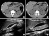
A 21-year-old man who had no medical history was admitted to the hospital due to lower abdominal pain that he had for 1 day. Physical examination revealed direct tenderness on the right lower quadrant without rebound tenderness. The laboratory evaluation was within the normal range except mild leukocytosis. Computed tomography (CT) showed cecal diverticulitis and there was no evidence of stone in the GB (Fig. 1A). Five days of intravenous ceftriaxone (2 g i.v. q 24 hours) was administered for controlling the diverticulitis, and consequently the pain was much improved. The follow-up CT, which was performed on the 8th hospital day, showed a newly developed huge GB stone without inflammation (Fig. 1B), and a 2.5 × 3.5 cm sized GB stone with an acoustic shadow was observed on ultrasonography (Fig. 1C). The patient had no symptoms to suspect cholecystitis, and the ceftriaxone had already been stopped, so we decided to observe this stone and the patient was discharged. Follow-up ultrasonography that was performed one month later showed that there was no stones remained in the GB (Fig. 1D).
Case 2
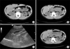
A 22-year-old man visited the emergency department due to fever for two days with aggravated dyspnea. Coarse breath sounds with rales were noted in both lungs, and a pneumonic infiltration was observed on the chest X-ray and chest CT. Twelve days of intravenous ceftriaxone (2 g i.v. q 24 hours) and seven days of clarythromycin (500 mg p.o. q 12 hours) was used to treat the pneumonia. Because the patient tolerated the antibiotics, he was not placed on bed rest and allowed to oral intake. The follow-up chest CT was performed on the 17th hospital day and it showed a newly developed stone in the GB (Fig. 2A, B). The ultrasonography that was performed on the 28th hospital day showed sustained GB sludge (Fig. 2C). The patient had no abdominal symptoms and the dyspnea and fever had resolved, so he was discharged on the 30th hospital day. The patient was readmitted to the hospital for follow-up and there was no more stone in the GB on the follow-up CT (Fig. 2D).
DISCUSSION
After usage of ceftriaxone, 60% of the drug is excreted into the urine and 40% of the drug is excreted into the bile in patients with normal renal function [4]. The mechanism of ceftriaxone associated GB sludge is as follows: the concentration of ceftriaxone in the GB can become 20 to 150 times greater than that in the serum, and excretion of ceftriaxone could disturb the excretion of the bile acids [5]. The concentration of ionized calcium in the bile is elevated and ceftriaxone can precipitate with calcium, like what occurs with bilirubin, and so ceftriaxone-associated biliary sludge is mainly composed of a calcium-ceftriaxone complex. Addition, ceftriaxone itself could affect the contractility of the GB [6].
The incidence of ceftriaxone-associated sludge or pseudolithiasis has been reported in 15 to 46% of the patients treated with ceftriaxone [1,7] in Korea, and the incidence was reported to be 30% (17 patients out of 57) of the pediatric patients who were treated with ceftriaxone. Sludge can also occur in the urinary tract [1], but only a few of these patients suffer from symptoms [7]. However, sometimes even gallstone pancreatitis or acute cholecystitis can occur after ceftriaxone treatment [8]. The main risk factors for ceftriaxone-associated GB sludge or stone was high daily dosages (over 2 g daily) and a long term duration of drug therapy [1,7]. Hypercalcemia, fasting and total parenteral nutrition, major surgery and dehydration can also cause this biliary sludge [9].
In general, biliary sludge can be expected to take 3 to 22 days after beginning ceftriaxone therapy [2,10] and it may reversible upon discontinuation of the drug with a range of 2 to 63 days after the end of treatment [2]. The sludge may serve as the nidus for gallstone pathogenesis, and the sludge commonly appears as a hypoechoic layer without acoustic shadowing. In our cases, we confirm that there was no GB stone initially, and GB stone was detected at 8th day and 17th day, respectively, after the ceftriaxone was started. The ceftriaxone-associated GB stone completely disappeared 1 month after discontinuation of the drug. There was GB sludge and no stone for the ultrasonographic findings of the 2nd case. This might be due to the interval between when this was first diagnosed via CT and the follow-up ultrasonography; the ceftriaxone had already been discontinued, so the stone may have resolved and became sludge.
In conclusion, GB stones can occur after ceftriaxone therapy and most of these resolved spontaneously after discontinuation of the drug. Clinicians, and especially surgeons, should aware of this complication and they should avoid unnecessary surgical interventions.
 XML Download