

 ePub
ePub Citation
 Print
Print


INTRODUCTION
Splenic infarction is a relatively uncommon diagnosis and this clinical presentation can mimic other causes of acute abdominal pain. Splenic infarction occurs as a consequence of systemic thromboembolism in association with several cardiovascular disorders [1]. We present a patient with celiac artery thromboembolism resulting in splenic infarction. The importance of this case, without any etiological predisposing factors, is that this kind of clinical situation should be considered in the differential diagnosis of acute abdomen.
CASE REPORT
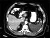
We present an unusual case of splenic infarction in a 53-year-old male without any etiological factors. A fifty-three-year-old male patient was admitted to our hospital with upper abdominal pain of sudden onset, which continued for 4-days, with complaints of nausea and vomiting. Abdominal rebound tenderness was found on physical examination, especially on the left upper quadrant. In laboratory examination; hemoglobin 10.6 g/dL, white blood cell 22,100/mm3, platelet 424,000/mm3, international normalized ratio 1.25 and activated partial thromboplastin time 31.20 s were determined. Acute embolism along common hepatic artery and splenic artery dilated proximal of celiac artery and multiple low-density wedge-shaped areas in the spleen, diagnosed as splenic infarction, were seen in abdominal computerized tomography (Fig. 1). This infarction area was segmental and 85% of total splenic volume. In digital subtraction angiography (DSA) imaging, acute thromboembolism proximal to the celiac artery and bifurcation of common hepatic and splenic artery were shown (Fig. 2). The distal hepatic artery was fed by gastroduodenal artery from a branch of the superior mesenteric artery. Thus, 80 IU/kg I.V. bolus and 18 IU/kg/hr infusion of heparin treatment was admini stered.
Transthoracic echocardiography and thorax high-resolution computed tomography were performed and thrombus was not detected in the heart or thoracic aorta. The risk factors for atherosclerosis including diabetes mellitus hypertension, hyperlipidemia and smoking were not determined in his past medical history and laboratory analysis. Furthermore, thrombus and/or atherosclerotic changes in descendent aorta and iliac vessels were not seen in DSA. Protein C, protein S and antithrombin III levels were normal. Anticardiolipin antibodies immunoglobulin (Ig) G and IgM and lupus anticoagulant were not determined. The homocysteine, MTFR, leiden and prothrombin gene mutation could not be found during the investigation of etiological factors. In 10 days of treatment, clinical findings and laboratory values had normalized. Warfarin therapy was ordered in the follow-up period and we determined re-canalization in celiac and splenic arteries with doppler ultrasonography after 6 months.
DISCUSSION
Splenic infarction is a relatively uncommon diagnosis. O'Keefe et al. [2] reviewed a large autopsy series and found that only 10% of splenic infarctions had been diagnosed ante mortem. Once a splenic infarction is identified, the source of the cause of infarction should be elicited such as hematologic, metabolic, or thromboembolic disease [3]. The etiology of thromboembolism is commonly cardiologic disorders including atrial fibrillation, ventricular aneurism and heart valve diseases [4]. Other etiological disorders can be listed as acute pancreatitis, antiphospholipid syndrome, malignancies, atherosclerosis, oral contraceptive drugs, diseases related with hypercoagulability and surgical trauma [5]. Pancreatitis is a common etiologic factor in patients with celiac artery thrombosis. Kumaran et al. [6] reported a case with isolated celiac trunk thrombosis caused by local complication of acute pancreatitis. They performed laparotomy due to clinical deterioration. Total gastrectomy and pancreatic debridement was carried out for visceral necrosis and the patient recovered well after six months. Serck and Cogbill [7] reported a 59-year-old female with sigmoid colon carcinoma had acute visceral ischemia originating from celiac trunk and superior mesenteric artery and, extensive visceral infarction ensued and she died. Also celiac and splenic arteries thrombus in a 14-year-old female who had received oral contraceptive agent for menorrhagia was reported by Arul et al. [8]. She was well at follow-up only with anticoagulant therapy and more aggressive therapy was not sought. Another cause of acute celiac occlusion is celiac artery dissection. Matsuo et al. [9] reported a 59-year-old male with isolated dissection of the celiac artery, which likely splenic infarction. They observed this patient without any treatment modalities because of the well developed collateral vessels. Furthermore, some authors applied transcatheter arterial embolization or surgical reconstruction because their patients became symptomatic with clinical deterioration in the follow-up period [10,11]. These treatment methods resulted in successful outcome in these patients.
An uncomplicated splenic infarction can be managed safely with medical treatment, but early surgical intervention is necessary when the complications of the infarct including abscess and rupture occurred [12]. Our case was diagnosed in the early period and anticoagulant therapy was performed in a short time so the result was successful. The importance of this case, without any etiological predisposing factors, is that this kind of clinical situation should be considered in the differential diagnosis of abdominal pain.

 XML Download