

 ePub
ePub Citation
 Print
Print


INTRODUCTION
Gastric cancer remains the second most common cancer diagnosed in the world. It accounts for 9.9% of all new cases of cancer diagnosed and is responsible for 12.1% of all cancer deaths [1]. Although the number of overall cancer deaths has declined, gastric cancer is still the leading cause of cancer death in Korea [2]. An essential step in managing gastric cancer is accurately assessing the preoperative stage and deciding on the adequate surgery including endoscopic treatment, minimal invasive surgery, and palliative operation.
However, managing gastric cancer is challenging because problems in determining treatment strategy remain as the prognosis has a wide range according to TNM classification [3]. There are limitations in the treatment of advanced gastric cancer (AGC) with surgery, whereas early gastric cancer (EGC) can be treated using minimal invasive surgery including endoscopy and laparoscopy with the limitation of lymphadenectomy [4,5]. Traditionally, computed tomography (CT) has been used for preoperative staging of gastric cancer. CT provides useful information on tumors based on anatomical structure, but there is a limitation in the accuracy of detection of EGC [6]. It was reported that the diagnostic accuracy in T staging can be increased using multi-detector row CT (MDCT), but N staging which is one of the most important prognostic factor as deciding treatment strategy for EGC remain unsatisfactory [7,8].
Recently, Positron emission tomography (PET) using 18F-fluorodeoxyglucose (18F-FDG) is being widely used to determine status of many different cancers, considered a new perspective on staging approach in malignancy. This advanced technology more accurately displays functional image of cancer with altered glucose metabolism, but lacks precision in localizing the tumor. A combined image of 18F-FDG-PET and CT (18F-FDG-PET/CT) can provide additional information by using the characteristics of both modalities. Improved staging accuracy has been demonstrated with the use of 18F-FDG-PET/CT in patients with lung and colon cancers [9-11].
18F-FDG-PET/CT has being widely used in Korea after the National Health Insurance Program decided to reimburse 18F-FDG-PET/CT cost in 2006 [12]. There is a paucity of data on the role of 18F-FDG-PET/CT in the preoperative diagnosis of gastric cancer. The purpose of this study is to compare the effectiveness of 18F-FDG-PET/CT to MDCT in terms of preoperative T and N staging of gastric cancer.
METHODS
Patients
A retrospective analysis of 78 preoperative 18F-FDG-PET/CT and MDCT in patients with gastric cancer who had undergone curative gastrectomy between February 2007 and October 2008 was performed. Informed consent for 18F-FDG-PET/CT and MDCT for the purpose of preoperative staging of gastric cancer was obtained from all patients. The patients comprised 53 men and 25 women with a median age of 61 years (range, 32 to 85 years). All of these patients underwent a preoperative staging procedure, including past history, physical examination, blood chemistry, abdominal MDCT, and esophagogastroduodenoscopy. 18F-FDG-PET/CT was performed within 4 weeks before gastrectomy.
MDCT technique
With MDCT unit, abdominal MDCT images were obtained. The MDCT scanner used was a 64-detector row scanner (Brilliance CT 64, Philips Medical System, Cleveland, OH, USA). All patients were given 4 g of effervescent granules (Top, Taejoon Pharmaceuticals, Seoul, Korea) to distend the stomach wall. The patients were placed in a supine position on the CT table. The acquisition volume included the whole abdomen from the dome of the diaphragm to lower margin of the symphysis pubis. An 18-gauge intravenous cannula was inserted into a vein in the antecubital fossa, forearm or wrist. Scanning of the abdomen was performed after intravenous injection with automatic power injector of 2 mL/kg of contrast medium (Ultravist 300, Schering, Berlin, Germany) at a flow rate of 2 to 3.5 mL/sec, total 120 mL. The scan delay time was determined by automatic bolus tracking method. The region of interest (ROI) was positioned at the descending aorta, at the level of the diaphragm. The hepatic arterial phase image was started 7 seconds after the attenuation reached 200 HU. Additional CT scan for portal phase images was started 60 seconds after the start of the contrast injection. The respective scanning parameters used for 16- and 64-MDCT scanners were 16 × 1.5 mm and 64 × 0.625 mm collimation, table feed of 24 and 46 mm per gantry rotation. X-ray tube voltage was 120 or 140 kVp, and amperage was determined by automatic dose reduction protocol. Volume data of MDCT scan were reconstructed as 5 mm thickness at 5 mm intervals. Coronal reformative images were reconstructed on a workstation, with a thickness of 3 mm at 3 mm intervals. All reconstructed images were sent to picture archiving and communication system (PACS) for evaluation.
18F-FDG-PET/CT technique
Patients were advised to fast for at least 6 hours prior to the intravenous injection of 5 to 6 MBq/kg of 18F-FDG. The 18F-FDG-PET/CT scan was obtained approximately 1 hour after administration of the FDG, using Biograph 6 system (Siemens, Knoxville, TN, USA). Seven or eight bed positions were imaged from the skull base to the mid thigh level. Patients were advised not to speak, chew, or move during scanning.
CT acquisition used the following parameters: 100 mA, 130 kVp tube voltage, helical thickness of 5 mm, and a helical pitch of 1.5:1. Using these parameters, the CT scan from the skull base to the mid thighs was obtained in 20 seconds. Immediately following the CT scan, a PET emission scan was acquired from the mid thigh to the base of skull with an acquisition time of 2.3 minutes per bed position based on body weight.
18F-FDG-PET/CT and MDCT image interpretation
An experienced gastrointestinal radiologist interpreted the MDCT images. Transverse and coronal MDCT images were evaluated with PACS monitors in random order. A lesion was determined to be cancerous when the gastric wall showed a focal thickening of at least 6 mm or greater or when focal enhancement was seen in the gastric wall. Lymph nodes were considered metastatic if they were large than 8 mm in the short-axis diameter and oval [13-15].
FDG uptake lesions of gastric wall and of lymph node bearing areas, regardless of size detected on PET/CT were regarded as malignancy, and the maximum standardized uptake value (SUVmax) was obtained using volume ROI.
Operation
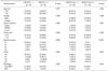
All patients underwent total gastrectomy (n = 19, 24.3%) or subtotal gastrectomy (n = 59, 75.7%) and standard lymphadenectomy (at least D2 lymphadenectomy). The advanced gastric cancer in 41 patients and EGC in 37 patients were revealed by pathologic diagnosis. According to the World Health Organization classification with Japanese modification [16], histopathological type of primary tumor was categorized as papillary adenocarcinoma (n = 1, 1.3%), well-differentiated adenocarcinoma (n = 4, 5.1%), moderately differentiated adenocarcinoma (n = 31, 39.7%), poorly differentiated adenocarcinoma (n = 22, 28.3%), mucinous adenocarcinoma (n = 3, 3.8%), and signet ring cell carcinoma (SRC) (n = 17, 21.8%).
Statistical analysis
We conducted Fisher's exact test for comparing the difference of respective categorical variables. The relation between SUVmax and pTNM stage was determined using Kruskall-Wallis test. The sensitivity and specificity of 18F-FDG-PET/CT and MDCT were calculated and compared using McNemar's chi-square test. The correlation analysis was performed to determine the relationship between tumor SUVmax and lymph node SUVmax, and also between tumor SUVmax and tumor size. P-values less than 0.05 were considered to be significant. The statistical analysis was carried out by the SPSS ver. 18.0 (SPSS Inc., Chicago, IL, USA).
RESULTS
Fifty one primary tumors were detected by preoperative 18F-FDG-PET/CT, and 27 patients had no noticeable results in 18F-FDG-PET/CT. Tumor size, depth of tumor, and lymph node metastasis (LNM) significantly affected the detection of primary tumor by 18F-FDG-PET/CT. As tumor size, depth of tumor, and LNM increased, the primary tumor was revealed more frequently by 18F-FDG-PET/CT. The detection rate of 18F-FDG-PET/CT for SRC was relatively lower than that for non-SRC (35.3% vs. 73.8%, P = 0.005). 18F-FDG-PET/CT detected LNM in 23 patients, and 55 patients showed no significant SUVmax of lymph node which was considered as metastasis. The results of 18F-FDG-PET/CT in detecting LNM were similar to the results of 18F-FDG-PET/CT in detecting primary tumor; however, tumor size had no association with detection of LNM on 18F-FDG-PET/CT (Table 1).
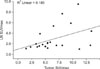
The correlation analysis showed a positive correlation exist between SUVmax of primary tumor and that of metastatic lymph node (P = 0.05, R = 0.43) (Fig. 1). However, there was no significant correlation between SUVmax of primary tumor and tumor size.
When assessed using the Kruskall-Wallis test, the mean SUVmax of primary tumor showed no significant difference between pT1, pT2, pT3, and pT4 (6.0 ± 1.4, 6.3 ± 0.6, 6.2 ± 0.8, and 5.4 ± 1.7). In addition, there was no statistical difference of the mean SUVmax of metastatic lymph node between pN0, pN1, pN2, and pN3 (1.8 ± 0.5, 3.1 ± 0.9, 5.6 ± 3.8, and 2.4 ± 0.5).
The detection rate of primary tumor by 18F-FDG-PET/CT and MDCT showed no significant difference. Among the 37 patients with EGC, 18F-FDG-PET/CT preoperatively detected 17 patients who were suspected to have gastric cancers. However, MDCT detected only 10 patients out of 37. The other 10 gastric cancer patients had no measurable lesions on both 18F-FDG-PET/CT and MDCT. A statistical difference between the detection rate of 18F-FDG-PET/CT and that of MDCT in patients with EGC was found (45.9% vs. 27.0%, P = 0.039). However, there was no significant difference between the detection rate of 18F-FDG-PET/CT (34/41 patients) and that of MDCT (37/41 patients) in AGC (Table 2). The SUVmax of primary tumor between EGC and AGC displayed similar result (6.0 ± 6.1, range 1.5 to 27.0 vs. 6.2 ± 2.9, range 2.2 to 13.2). However, the result of 18F-FDG-PET/CT (17 of 78 patients) for LNM confirmed pathologically was not significantly different from that of MDCT (23 of 78 patients) with respect to the detection of LNM (21.8% vs. 29.5%, P = 0.07). In 41 patients with AGC, MDCT revealed 22 LNM compared to 17 LNM on FDG-PET/CT (53.7% vs. 39.0%, P = 0.125). In EGC, detection rate of 18F-FDG-PET/CT (0 of 37 patients) for LNM did not show significant difference from that of MDCT (1 of 37 patients).
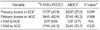
Overall, the accuracy of 18F-FDG-PET/CT was similar to that of MDCT in the diagnosis of LNM in gastric cancer (71.8% vs. 69.2%). However, the sensitivity of MDCT was superior to that of 18F-FDG-PET/CT (69.7% vs. 51.5%, P = 0.035). 18F-FDG-PET/CT was also superior to MDCT in terms of the specificity (86.7% vs. 75.6%, P = 0.029) (Table 3).
DISCUSSION
MDCT has also been widely used in the preoperative diagnosis of gastric cancer, providing high-resolution anatomic details about staging, but is limited by the use of size criteria [17]. FDG-PET lacks accurate anatomical orientation when displaying metabolic information of malignancy [18]. The combination of PET and CT has emerged as the most accurate technology and provides good anatomical localization of functional data [19].
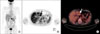
18F-FDG-PET/CT is increasingly used in the preoperative diagnosis of various cancers to determine staging, as well as in the detection of recurrence after curative surgery [9]. And recently, for the purpose of the detection of recurrence for gastric cancer after curative gastrectomy, 18F-FDG-PET/CT has been introduced and shown comparable results to CT [20]. As shown in this study, the performance of 18F-FDG-PET/CT was comparable to that of MDCT with a similar 71.8% accuracy. The interesting result of 18F-FDG-PET/CT over MDCT in this study was an improvement in the detection of EGC (45.9% vs. 27.0%), while there was no significant difference of SUVmax between EGC and AGC (6.0 ± 6.1 vs. 6.2 ± 2.9). The metabolic alteration of tumor allowed 18F-FDG-PET/CT to detect lesion regardless of tumor advancement (Fig. 2).
In the present study, the detection rate of primary tumor and LNM in 18F-FDG-PET/CT was significantly higher as the T and N stage increased, while the tumor size was not significantly associated with the detection of LNM in 18F-FDG-PET/CT. The histological subtype had a strong influence on the detection of primary tumor and LNM. The detection rate of SRC in 18F-FDG-PET/CT was lower than that of non-SRC in terms of detection of primary tumor and LNM. The FDG uptake in primary tumor and LNM in cases of signet ring cell carcinoma was less pronounced than that in non-SRC [21,22]. The results suggest that 18F-FDG-PET/CT appears to play a less useful diagnostic role in such cases.
The evaluation of 18F-FDG-PET/CT compared to MDCT in the diagnostic accuracy of EGC has not achieved consistent levels of accuracy. Previous reports have shown low detection rates for EGC in CT, with a range of 26 to 53% [23,24]. In one study, the detection rate of mucosal cancer was very low (16.7%) in comparison to that for submucosal cancer (68.8%) in MDCT [25]. In the present study, the detection rate of mucosal cancer and submucosal cancer in 18F-FDG-PET/CT was 35.0% (7/20) and 58.8% (10/17), respectively, which is considerably higher detection rate for cases of mucosal cancer. From this result, 18F-FDG-PET/CT displays greater superiority with respect to the detection of mucosal gastric cancer compared to MDCT, but has a limited role compared to other modalities [26].
We found a borderline statistical significant positive correlation in SUVmax between the primary tumor and lymph node. The high SUVmax of primary tumor is associated with better detection rate for LNM. High SUVmax in the primary tumor had a positive impact on the accuracy of 18F-FDG-PET/CT assessment of LNM. Gastric cancer with SRC tended to be associated with a lower detection rate in both the primary tumor and LNM in 18F-FDG-PET/CT, and 18F-FDG-PET/CT may not be sufficient for preoperative staging in gastric cancer with SRC.
FDG-PET has a low to moderate sensitivity of LNM due to its limited resolution; FDG-PET have a 4- to 5-mm resolution [27], but 14.5% of metastatic lymph node in gastric cancer has the largest diameter of less than 3 mm [28]. Another reason for the low sensitivity of FDG-PET is the masking of perigastric lymph node by the FDG uptake of the adjacent primary tumor. 18F-FDG-PET/CT provides both anatomical and functional information and displays more accurate localization of both the primary tumor and lymph node with increased SUV than FDG-PET alone. We found that the sensitivity and specificity of 18F-FDG-PET/CT in this study was comparable to a previous report in assessing lymph node status in gastric cancer (51.5% and 86.7% vs. 54.7% and 92.2%) [29].
The interesting finding from the present study was that both techniques had important informative role in preoperative staging of gastric cancer. 18F-FDG-PET/CT was superior to MDCT in terms of the detection of the primary tumor in EGC, while MDCT provided more accuracy of LNM than 18F-FDG-PET/CT in AGC.
In conclusion, 18F-FDG-PET/CT has a better diagnostic performance than MDCT in detecting a primary tumor in EGC and comparable accuracy of detecting LNM. However, 18F-FDG-PET/CT shows no definite improvement over MDCT in detecting primary tumors and LNM in patients with gastric cancer. Moreover, 18F-FDG-PET/CT has clear limitations in preoperative staging in case of patients with SRC. This limitation must be taken into consideration when assessing gastric cancer. Therefore, randomized controlled trial on the application of 18F-FDG-PET/CT in the diagnosis of EGC and AGC, respectively, is advocated.
 XML Download