

 ePub
ePub Citation
 Print
Print


INTRODUCTION
In addition to the age of patients and the total body surface area burned, inhalation injury has been known as a factor that seriously affects a prognosis of patients with burn [1,2]. Its incidence has been reported to vary, but it generally occurs in approximately 7 to 20% of patients with burn who are in need of hospitalization [3]. Besides, it has also been reported that inhalation burn injury is concurrently present in 30% of patients with severe burn. According to some reports, it increases the mortality by 20% and the incidence of pneumonia by 40% [4].
The inhalation burn injury causes the decreased oxygen perfusion due to the direct injuries to the upper and lower respiratory tracts, alveolar injury induced by mechanical ventilation, secondary pneumonia and acute respiratory distress syndrome. Thus, it has been reported to have a close relationship with a prognosis of the burn patients. Accordingly, an accurate diagnosis and treatment of the inhalation burn injury in the early stage would be essential for achieving a good prognosis of patients with burn.
But the inhalation burn injury has a latent period for 3 to 4 days prior to the occurrence of respiratory complications [5]. Until 1 to 2 days after the onset of inhalation burn injury, abnormal findings cannot be identified on a chest radiography or an arterial blood gas analysis [6,7]. An early diagnosis of inhalation burn injury cannot easily be made based on its characteristics described above. In the past, a diagnosis of inhalation burn injury was made based on the clinical signs or a past medical history of patients. At the present, however, a bronchoscopy is established as a standard treatment modality for the treatment of patients with inhalation burn injury. As compared with the past, the degree of diagnostic accuracy has been improved [8]. Thereafter, many studies have been conducted to examine the inhalation burn injury. It has been reported that the findings seen on a bronchoscopy are useful in predicting the progression of acute lung injury [9]. In an actual clinical setting, however, there are often cases in which the findings seen on a bronchoscopy are not in agreement with the severity of respiratory complication and the prognosis of patients.
In the current study, following an analysis of the incidence and mortality of inhalation burn injury in Korea, their effects on a prognosis of patients were evaluated. In addition, statistical analysis was performed to identify the correlations between the representative indicators that are used to assess the inhalation burn injury, such as PaO2/FiO2 (P/F) ratio, bronchoscopic findings and carboxyhemoglobin (COHb) level, and a prognosis of patients. Thus, attempts were made to assess the significance of these indicators as a prognostic factor for patients with inhalation burn injury.
METHODS
Of patients who were hospitalized at a burn intensive care unit (ICU) of Hangang Sacred Heart Hospital of Hallym University College of Medicine during a period ranging from January 1, 2008 to December 31, 2009, those who were suspected to have an inhalation injury and then underwent a bronchoscopy were enrolled in the current study. Of these, excluding patients who died of burn shock within three days following hospitalization, those who admitted to burn ICU 12 hours after burn injury and those who underwent a bronchoscopy 48 hours after injury, 170 were enrolled in the current retrospective study.
In all the patients, COHb was measured through a blood sampling immediately after they visited an emergency care center. The P/F ratio was measured by arterial blood gas analysis at 24 hours after burn injury when the initial fluid therapy was completed. For the pre-treatment for a bronchoscopy, patients were given an intramuscular injection of atropine 0.5 mg and pethidine 25 to 39 mg 30 minutes before. For local anesthesia, patients were inhaled 4% lidocaine 5 mL (200 mg) through a nasal spray for 10 to 15 minutes. Bronchoscopy was performed through oral or nasal insertion of bronchoscope. For the assessment of the bronchoscopic findings, eight parameters were evaluated and these include edema, blistering, carbonaceous material, soot, hemorrhage, inflammation, ulceration and necrosis of mucosa. Of these, cases corresponding to 1 to 3 parameters, 4 to 6 parameters and 7 to 8 parameters were classified as the mild, moderate and severe types, respectively. Statistical analysis was performed with a chi-square test, for which a statistical software package, SPSS ver. 13.0 (SPSS Inc., Chicago, IL, USA), was used.
RESULTS
Demographics of the patients
Cause of injury of 170 investigated patients was all flame burn. Mean age of patients was 45 ± 13 years (range, 11 to 90 years). Patients were composed of 135 men and 35 women, where the male-to-female ratio was 3.8:1. Mean percentage of total body surface area (TBSA) burned was 43.9 ± 26.2%.
The incidence and mortality of patients with inhalation injury
Of 170 study patients, 28 patients no bronchoscopic findings that are suggestive of inhalation burn injury and 142 patients had a diagnosis of inhalation injury confirmed by bronchoscopy. During the study period, the number of patients admitted to our burn ICU was 671. So the incidence of inhalation injury of our burn ICU patients was 21.1%. In 142 patients with inhalation burn injury, the mean percentage of burn was 44.6 ± 26.7%. The overall mortality of patients with inhalation burn injury was 35.2%.
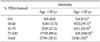
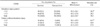
Following the classification of patients based on the mean percentage of burn, there was a significant correlation between the percentage of burn and the mortality (P = 0.000). Besides, following the classification of patients based on the age, the mortality was found to be 28.1% in patients aged 50 years or younger and 50% in those aged 50 years or older. This difference reached a statistical significance (P = 0.018). In the patients whose TBSA burned was between 10% and 70%, mortality of the patients younger than 50 was significantly lower than the patients older than 50. But when the percentage of TBSA burned was under 9% or over 70%, there was no significant differences of mortality according to patients' age (Table 1).
A comparison of the mortality between patients with inhalation burn injury and the total hospitalized patients
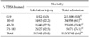
There were no deaths in those whose percentage of burn was under 9%. Of 130 patients with inhalation injury whose percentage of burn was over 10%, 50 patients died and the mortality was 38.4%. Based on the percentage of TBSA burned, the mortality was 22.2% in patients whose percentage of burn was between 10% and 40%, 27.5% in those whose percentage of burn was between 41% and 70% and 92.5% in those whose percentage of burn was between 71% and 100%. These results indicate that the mortality was increased as the percentage of burn was increased, which also reached a statistical significance (P = 0.000).
Following the classification of total patients who admitted to our burn ICU during the investigation period, the mortality was found to be 6.1% in patients whose percentage of burn was 10 to 40%, 23.8% in those whose percentage of burn was 41 to 70% and 76.1% in those whose percentage of burn was 71 to 100%. Mortality rate of total patients was significantly increased as the percentage of TBSA burned was increased (P = 0.000) (Table 2).
When compared the mortality between patients with inhalation injury and total burn ICU patients during investigation period, there was no significant difference in the mortality between the two groups if the percentage of TBSA burned was under 9% or over 40%. But, in the percentage of TBSA burned between 10% and 40%, the mortality was significantly higher in patients with inhalation injury as compared with total patients (P = 0.000).
Findings of bronchoscopy within 48 hours from injury
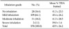
Bronchoscopic findings were evaluated with eight parameters. In 142 patients with inhalation injury, all the patients had the finding of edema. And, there were 19 cases of blister, 51 cases of soot, 7 cases of hemorrhage, 1 case of necrosis of mucosa, 6 cases of ulceration, 6 cases of inflammation and 18 cases of carbonaceous material. So there were 109 cases of mild inhalation, 31 cases of moderate inhalation and two cases of severe inhalation (Table 3).
The mortality of patients by bronchoscopic result within 48 hours from injury
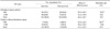
Twenty-eight patients were declared not to have inhalation injury. In these patients, mean percentage of TBSA burned was 41.3 ± 23.3% and the mortality was 17.8%. In 142 patients who had a diagnosis of inhalation injury confirmed on bronchoscopy, mean percentage of TBSA burned was 44.4 ± 26.7%. Of these, 50 patients died. So mortality of patients with inhalation injury was 35.2%. It is somewhat regrettable that a comparison was not made following the classification of patients based on the severity inhalation burn injury. Because there were a relatively greater number of patients with mild inhalation and only two patients were determined to be severe cases, however, the moderate and severe cases were classified into a single group for a comparison of the mortality. Of total patients with inhalation burn injury, based on the bronchoscopic findings, mean percentage of TBSA burned was 45.4 ± 26.2% and the mortality was 31.1% in mild cases. In the moderate and severe cases, mean percentage of TBSA burned was 40.0 ± 28.1% and the mortality was 48.5%. There was a significant difference in the mortality between the three groups (P = 0.035) (Table 4).
The mortality of patients by P/F ratio after 24 hours from injury
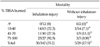
Of 142 inhalation patients, mean percentage of TBSA burned was 55.5 ± 26.3% and the mean mortality rate was 53.8% in 78 patients whose P/F ratio was less than 300. In 64 patients whose P/F ratio was more than 300, mean percentage of TBSA burned was 31.2 ± 19.0% and the mean mortality rate was 12.5%. There was a significant difference in the mortality between the two groups (P = 0.000) (Table 5).
Of 28 patients who had no inhalation injury on bronchoscopy, mean percentage of TBSA burned was 52.0 ± 23.3% and the mean mortality rate was 36.3% in 11 patients whose P/F ratio was less than 300. In 17 patients whose P/F ratio was more than 300, mean percentage of TBSA burned was 34.4 ± 22.2% and the mean mortality rate was 0.06%. The mortality was higher in the group where the P/F ratio was less than 300. But there was no significant difference in the mortality between the two groups (P = 0.062) (Table 5).
The mortality of patients by the COHb
In 142 patients with inhalation injury, mean percentage of TBSA burned was 45.3 ± 26.1% and the mean mortality was 34.2% in 73 patients whose COHb was < 1.5. Besides, mean percentage of TBSA burned was 43.2 ± 27.4% and the mean mortality was 36.2% in 69 patients whose COHb was > 1.6. Based on the COHb level, there was no significant difference in the mortality between the two groups (P = 0.943) (Table 6).
In 28 patients who had no inhalation injury on bronchoscopy, mean percentage of TBSA burned was 38.1 ± 25.3% and the mean mortality was 16.7% in patients whose COHb was <1.5. Besides, mean percentage of TBSA burned was 43.7 ± 23.4% and the mean mortality was 18.8% in 16 patients whose COHb was >1.6. There was no significant difference in the mortality between the two groups (P = 1.000) (Table 6).
A comparison of the P/F ratio and COHb between the group with inhalation injury and the group without inhalation injury
In 142 patients who had a diagnosis of inhalation injury confirmed by bronchoscopy, the mean P/F ratio was 294.7 ± 109.1 and the mean COHb was 3.0 ± 4.1. In 28 patients who had no findings of inhalation injury on bronchoscopy, the mean P/F ratio was 341.5 ± 99.8 and the mean COHb was 1.9 ± 1.1. There were significant differences in the P/F ratio and COHb between the two groups (Table 7).
DISCUSSION
Necessity of the early assessment and diagnosis of inhalation injury with bronchoscopy is still in controversy. Bronchoscopy is considered as a standard test regimen for all the patients who are suspected to have an inhalation injury. This is because the testing cost is not relatively higher; a prompt test can be performed by experienced personnel; and it has usefulness in evaluating the damage of the respiratory tract. A bronchoscopy is useful in screening the bronchial edema, erythema or carbon deposition, but it cannot have an absolute effect on the treatment of smoke inhalation. Bronchoscopy has the limitation that the bronchoscopic results depend on the investigators. So a thorough history taking, physical examination and laboratory results should be concomitantly used to make a diagnosis of inhalation injury. The therapeutic use of a bronchoscope would be of significance to the treatment of patients if it is applied for the lobar expansion in the atelectatic lung or the removal of the intrabronchial secretions or lumps obstructing the airway tract.
Edelman et al. [3] reported that 229 of 829 patients had a diagnosis of inhalation injury confirmed and its incidence was 28% accordingly. These authors reported that the mortality of 76 patients with inhalation injury was 16%. In addition the mortality was 3% in patients with percentage of TBSA burned of 1 to 9%, 12% in those with percentage of TBSA burned of 10 to 29%, 33% in those with percentage of TBSA burned of 30 to 49% and 42% in those with percentage of TBSA burned of >50%. Besides, these authors also reported that the mortality was 23% in patients with inhalation burn injury who had a percentage of burn of >10%. Then, they noted that the mortality was significantly higher as compared with patients with inhalation injury whose percentage of TBSA burned was less than 10% and those who solely had an inhalation injury without burn. There was no significant difference in the mortality, however, between patients with inhalation burn injury who had a percentage of TBSA burned of <10% and those who solely had an inhalation burn injury. Because there were only 19 patients whose percentage of TBSA burned was >50%, however, no statistical analysis could be performed for patients with massive burns.
Mortality rate of total patients who admitted to our burn ICU during the investigation period was compared with the mortality rate of patients with inhalation injury. In patients whose percentage of TBSA burned was <9% or >40%, there was no significant difference in the mortality between inhalation injury patients and total patients. In patients whose percentage of TBSA burned was 10 to 40%, the mortality rate of inhalaton injury patients was significantly higher when compared with the total patients (Table 2).
Edelman et al. [3] reported that there was a significant correlation between the age and the mortality. In patients with inhalation injury whose percentage of TBSA burned was <10%, there was no significant difference in the mortality between the group of 50 years or older and the group less than 50 years. In patients with inhalation injury whose percentage of TBSA burned was 10 to 49%, however, there was a significant difference in the mortality between the group of 50 years or older and the group less than 50 years. In patients with percentage of TBSA burned over 50%, no statistical analysis could be performed due to a small number of enrolled patients.
In our study, the age-related difference in the mortality depending on the size of burn injury was evaluated in patients with inhalation injury. According to this, in patients with percentage of TBSA burned between 10% and 70%, there was significant difference in the mortality between the group of 50 years or older and the group less than 50 years. In patients with percentage of TBSA burned less than 9% or more than 70%, however, there was significant difference in the mortality between the two age groups (Table 1). In association with these results, in patients with percentage of TBSA burned more than 70%, the mortality was 92.5%. The burn injury itself has a great impact on a prognosis of patients. In patients with percentage of TBSA burned less than 70%, however, the mortality was markedly decreased to 27.5%. It can therefore be inferred that the age has a greater impact on a prognosis of patients from a relative perspective.
According to Finnerty et al. [10], in pediatric patients with severe burn injury whose percentage of TBSA burned was more than 60%, the mortality rate was 40% in the group with inhalation injury and 12% without it. Then, the authors reported that the mortality was significantly higher in the group where there was a concurrent presence of inhalation injury.
In our clinical series of patients, however, in patients whose percentage of TBSA burned was between 10% and 40%, the mortality was significantly higher in those with inhalation injury as compared with total patients. In patients with percentage of TBSA burned more than 40%, the mortality was higher in those with inhalation injury as compared with total patients. But there was no statistical significance.
Aside from the above results, following a comparison of the mortality between 28 patients who had no inhalation injury finding on bronchoscopy and 142 patients who were diagnosed with inhalation injury, there were no significant differences in the mortality rate in all groups classified by burn size (Table 8).
Hassan et al. [9] examined the mortality in 105 patients with inhalation injury following the classification based on the severity on bronchoscopy. According to these authors, the mortality rate was 18% in patients who had no inhalation injury, 25.5% in those who had mild inhalation injury, 40.6% in those who had moderate inhalation injury and 66.7% in those who had severe inhalation injury. Also in the current study, following the classification of patients based on a bronchoscop into patients who had no inhalation injury, those who had mild inhalation injury, those who had moderate inhalation burn injury and those who had severe inhalation injury, the mortality was found to be 17.8%, 31.1% and 48.5% in the corresponding order. These results indicate that there was a significant correlation between the findings of bronchoscopy and the mortality (Table 4).
In our study, in total of 142 patients with inhalation injury, 109 patients were classified as mild cases and only two were classified as severe cases. To this extent, there were smaller number of patients with severe inhalation injury. This might be due to a greater number of patients classified as severe cases when they were determined to have more than seven of eight parameters, such as edema, blistering, carbonaceous material, soot, hemorrhage, inflammation, ulceration and necrosis of mucosa, because we had strict criteria for bronchoscopic findings at our medical institution. As a matter of fact, of 142 patients with inhalation burn injury, there were only seven cases of hemorrhage, six cases of ulceration, six cases of inflammation and one case of necrosis of mucosa.
In reporting bronchoscopic findings, Hassan et al. [9] classified the severity of inhalation injury based on the shape of mucosa. According to these authors, the mild cases include mild erythema; the moderate cases include erythema and swelling; and the severe cases include pale, dry and necrosis. Besides, according to Chou et al. [6], based on the findings of a bronchoscopy, the mild cases include mild edema and hyperemia with without carbon soot; the moderate cases include severe edema and hyperemia with or without carbon soot; and the severe cases include ulceration, necrosis, absence of both cough reflex and bronchial secretions. These authors also reported that the mortality was 4% in mild cases, 33% in moderate cases and 77% in severe cases, based on which they noted that there was a significant correlation between the findings of a bronchoscopy and the mortality.
There are no international standards for the classification of bronchoscopic findings in patients with inhalation injury. Because bronchoscopic findings are determined subjectively by examiner, there can be a discrepancy between bronchoscopic findings and the clinical symptoms and the mortality of the inhalation injury patients are reported variously. But most of the articles about inhalation injury reports that the greater the severity of inhalation injury was, the higher the mortality became as shown in our results.
When investigate COHb level and the P/F ratio of inhalation injury patients and the patients without inhalation injury, mean P/F ratio and the mean COHb level of inhalation injury patients were 294.7 ± 109.1 and 3.0 ± 4.1. These results were significantly higher than those seen in patients without inhalation injury.
The P/F ratio is a ratio of the partial pressure of arterial blood oxygen to the concentration of inhaled oxygen and it is a relative indicator for hypoxia. A lower level of P/F ratio is indicative of the diffusion disorder, the ventilation disorder, a lack of the consistency between ventilation and perfusion and true shunt. According to Brown et al. [11], the survival rate was increased with the patients whose P/F ratio were over 300 when they analyzed the P/F ratio at the time of the completion of fluid therapy in 120 patients with inhalation injury. In the current study, 142 inhalation injury patients were classified into two groups with a P/F ratio of 300. The mortality rate of patients whose P/F ratio were over 300 was 12.5% and 53.8% in patients whose P/F ratio were under 300. This results reached to the statistical significance (Table 5). According to Hassan et al. [9], except for the group without inhalation injury on bronchoscopy, with increasing the severity of inhalation injury, P/F ratio of patients were decreased.
Traditionally it has been known that the COHb level is an indicator of inhalation injury. However, the COHb level can be elevated up to 8% in smokers and 10% in the residents of air pollution. A half-life of COHb is 3 to 4 hours indoors and it becomes shorter at a high partial pressure of oxygen. According to Hassan et al. [9], based on the severity of inhalation injury on bronchoscopy, the mean COHb level was 5.54 in the mild cases, 20.67 in the moderate cases and 20.32 in the severe cases. These differences reached a statistical significance. In our study, the mean COHb level was 1.9 ± 1.2 in patients who had no inhalation burn injury, 1.9 ± 1.8 in those who had mild inhalation injury and 6.7 ± 6.9 in moderate and severe inhalation burn injury. These differences reached a statistical significance. In regard to the correlation between COHb level and the mortality, a comparison of the mortality was made based on a COHb level of 1.5%, the normal COHb level seen in non-smokers, between patients with inhalation injury and patients without inhalation injury. In both groups, there was no significant correlation between COHb level and the mortality (Table 6). This might be because our study was conducted in patients who arrived at our hospital within 12 hours following the onset of injury, and a mean length of time of 5.96 ± 3.64 hours elapsed until patients were transferred to us, exceeding a half-life, and most of the patients inhaled oxygen during transfer.
In conclusion, it has been reported that the presence of inhalation injury is closely associated with a prognosis of patients in addition to the percentage of TBSA burned and the age of patients. In our study, in patients with percentage of TBSA burned of 10 to 40%, the mortality was significantly higher in patients with inhalation burn injury as compared with the total hospitalized patients. In patients with percentage of TBSA burned less than 10% or more than 40%, however, there was no significant difference in the mortality between patients with inhalation injury and the total hospitalized patients. It was also shown that the mortality of patients was increased as the severity of inhalation injury based on bronchoscopic finding was increased. Furthermore, bronchoscopic finding and the P/F ratio at the time of the completion of fluid therapy were found to have a correlation with a prognosis of patients with inhalation burn injury. But the COHb level was found to have no correlation with a prognosis of patients with inhalation burn injury.
Bronchoscopy is currently a standard diagnostic regimen for inhalation injury. But there are no international standard criteria for determining bronchoscopic findings, for which further studies and international collaborative approaches might be mandatory. With the introduction of a well-organized evaluation system for inhalation injury, an objective classification of patients with inhalation burn injury would become possible. This might greatly contribute to the treatment of patients and research.


 XML Download