

 Citation
 Print
Print


INTRODUCTION
Adventitial cystic disease (ACD) is a well-characterized vascular disease by a vessel wall based cystic lesion with mucinous contents. Adventitial cystic disease commonly develops in the arteries. Venous ACD is extremely rare with fewer than 20 cases reported in the world-wide literature [1]. We report the cases of one 40 and one 68-year-old woman with ACD arising in the common femoral vein who were successfully treated surgically.
CASE REPORTS
Case 1
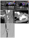
A 40-year-old woman presented with a 10-day history of left lower extremity painless swelling. She had no previous history of trauma or malignancy. On examination, her left leg was obviously swollen with no tenderness, and no palpable masses or lymphadenopathy. A venous ultrasound examination was performed, the results of which reported a typical hypoechoic fluid filled cystic structure in the femoral vein causing a mass effect on the adjacent common femoral vein lumen (Fig. 1). Computed tomography (CT) and phlebography of the left leg revealed that a short segment of the common femoral vein was nearly obliterated by an unenhanced mass (Fig. 1). From this information we diagnosed ACD in the common femoral vein and decided on surgical correction. At surgery the common femoral artery was exposed and retracted laterally to facilitate dissection of the common femoral vein. The common femoral vein, profunda femoris, and superficial femoral veins were dissected and controlled, revealing an enlarged common femoral vein with a bluish color (Fig. 2). The lumen was almost entirely compressed by a large unilocular, intramural cystic structure in the posterior wall of the vein. A longitudinal venotomy was made in the posterior wall to reveal thick gelatinous mucoid material lying within a cystic cavity (Fig. 2). The evacuation of gelatin material was performed along the entire length of cyst by squeezing and then the excision of the posterior cystic wall was performed. Flow in the common femoral vein was resumed after evacuation. Postoperatively, the patient received anticoagulation with warfarin and made an uneventful recovery. At the 6-month follow-up, the swelling in the leg had resolved, and the common femoral vein was patent on color duplex imaging, with no mass effect or recurrence.
Case 2
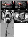
A 68-year-old woman presented with a one-month history of unexplained edema of the right lower extremity. On physical examination, her right leg was swollen with no tenderness, and no palpable masses. She had no previous history of trauma, right leg operation or malignancy. A venous duplex scan was performed. The results showed a hypoechoic fluid filled cyst in right common femoral vein and luminal narrowing of the adjacent vein (Fig. 3). CT and phlebography showed focal segmental obliteration of the common femoral vein by extrinsic mass (Fig. 3). We also confirmed ACD in the common femoral vein and performed surgical exploration. The lumen of the common femoral vein was almost entirely compressed by a large unilocular, intramural cystic structure in the lateral wall of the vein. A longitudinal venotomy was made in the lateral posterior wall to reveal thick gelatinous mucoid material lying within a cystic cavity (Fig. 4). The excision of the adventitial wall was performed to restore normal blood flow (Fig. 4). The patient was released after short hospitalization and received anticoagulation with warfarin for 6 months. At 6-month follow-up the patient remained symptom free and without evidence of recurrence on the venous duplex scan.
DISCUSSION
Adventitial cystic disease is a well-described pathology of the popliteal artery but is rarely reported in veins. In 1998, Levien and Benn [2] identified 323 confirmed cases of ACD of the blood vessels, only 17 (5.3%) of which involved a vein. Reported venous ACD is commonly seen in middle-aged men. According to the review of 18 cases of ACD involving the venous system, ACD of the vein tends to develop later in life, at an average age of 45.4 years (range, 23 to 75 years). Among the 18 patients, 11 were men and 7 were women (M:F ratio, 1.6:1). The affected veins were the femoral (9 cases), popliteal (3 cases), small saphenous (2 cases), iliac (2 cases), great saphenous (1 case), and wrist (1 case) [3]. Predominant presenting symptoms are extremity swelling and groin mass. The clinical presentation may mimic DVT and the differential diagnosis including femoral aneurysm, ganglion cyst, lipoma, venous leiomyoma, malignancy, or lymphadenopathy is needed.
On the other hand, arterial ACD is most commonly encountered in the popliteal artery, but also reported in the iliac, femoral, radial, and ulnar arteries. Arterial ACD is often included on the differential list for claudication symptoms in young men without risk factors. It is estimated to account for 0.1% of all patients who seek treatment for claudication. The epidemiology of arterial ACD finds a male/female ratio of approximately 15:1 and an average age in the fourth to the fifth decade [4].
The etiology of ACD is not well understood, although various theories have been proposed. Repeated microtrauma, ectopic aganglionosis, degeneration of the adventitia due to connective tissue diseases and developmental theory have been discussed [5]. However, most theories on etiology focus on arterial ACD, which might not be relevant to venous ACD. There are several differences between arterial and venous ACD. Thus, it is hard to define a common theory for the development of ACD. The differences of arterial and venous ACD are followings; (1) that no cases of joint-associated venous ACD have been reported, (2) that the much higher male preponderance of arterial ACD than venous ACD [4], (3) that venous ACD rarely affects the popliteal segment, which is most common in arterial ACD, (4) and that thers is a large difference in incidence, with arterial ACD estimated at 1:1,200 patients with claudication, but only countable numbers of venous ACDs have been reported [3].
Diagnostic tests may include venous duplex scan, CT scan or magnetic resonance imaging (MRI). Duplex scan may show a vein wall associated hypoechoic fluid-filled cyst with a posterior acoustic window, which may provide specific diagnostic clues to decision making. CT scan or MRI tests may reveal an extraluminal mass in close proximity to the vein. Ascending venography may show a luminal defect from external compression, which was present in this case.
Minimally invasive management has been reported with image-guided needle aspiration of adventitial cysts, but incomplete evacuation of cysts secondary to high viscosity and the mucin-secreting mesenchymal cells that are left in situ have resulted in high recurrence rates [6]. Therefore, we believe surgical intervention should be the treatment of choice. The preferred surgical management of adventitial cystic disease is either transadventitial or- transluminal evacuation of the mucoid cysts with or without removal of cystic wall, as is in the cases we have described. Surgical treatments for lesions in the femoral vein also include transluminal fenestration and segmental resection. Resection with grafting or a patch angioplasty is necessary when the vessel is occluded [1]. But, the long-term results of surgical treatment are unknown till now because of its rarity. Endovascular treatment is ineffective because it fails to deal with the underlying cause of compression and may become problematic with stents crossing joint lines and with thrombosis in low flow veins [5].
ACD is rare, but should be suspected in patients presenting with spontaneous first-time leg swelling, especially where the studies suggest an extrinsic mass. The proper treatment is open surgical management.


 XML Download