

 ePub
ePub Citation
 Print
Print


INTRODUCTION
Persistent cloaca is one of the most severe types of anorectal malformations occurring in newborn infants. Appropriate drainage is difficult because the degree of malformation differs among individuals and difficulty to determine the exact anatomical structure in each patient due to hydrocolpos or dilated urinary bladder and colon. Later reconstruction is also dependent on individual characteristics and type of the surgical procedure performed. We describe here our experiences with surgical management of the children with persistent cloaca.
METHODS
Children diagnosed with persistent cloaca at the Asan Medical Center from 1998 to 2010 were retrospectively identified, and their clinical histories and radiological and surgical findings were analyzed.
RESULTS
Clinical features
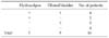
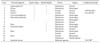
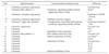
Sixteen patients were managed in their neonatal period (Table 1). Eleven infants prenatally diagnosed as following abnormalities, 3 were persistent cloaca, 8 were other disease (Tables 2, 3). Associate anomalies were combined in 3 patients, 1 patient combined the vertebral, anorectal, cardiac, esophageal atresia with tracheoesophageal fistula, renal and radial and limb anomalies (VACTER) syndrome. Chromosomal spreads were performed in 9 patients, with 1 having a 47.XX +marker chromosome. The father of this patient had the same chromosomal abnormality, as well as a surgical history of imperforate anus.
Management in neonatal period
In 4 patients, hydrocolpos and dilated bladder were detected at the time of birth. In 3 of these patients, cystostomy and vaginostomy were performed immediately after birth, they had no further problems other than urinary tract infections. One patient (no.2) with a foley catheter during 2 months after birth underwent cystostomy and vaginostomy due to persistent extension of the vagina and bladder.
Three patients had hydrocolpos only without any bladder enlargement. One patient (No.5) who did not undergo vaginostomy initially underwent a cystostomy and vaginostomy at 30th day after birth, but died of ventriculitis and hydrocephalus 6 months later due to sepsis caused by urinary tract infection. One patient (No.6) who underwent a vaginostomy had no further problems for 4 months. Another patient (No.7) was underwent a colostomy at another hospital did not require further treatment.
Dilated bladder without hydrocolpos was detected in 5 patients. In one patient (No.12) of these, a cystostomy was performed 18 days after birth, because urine was drained via a colostomy. Intermittent urinary drainage was performed through a colostomy in 1 patient (No.8) and internal urethrotomy was performed in another 1 patient (No.10) with stenosis of the bladder neck. The remaining 2 patients did not require any other procedures.
The remaining 4 patients did not show enlargement of the vagina and bladder. Three patients of these did not require any urinary or vaginal catheter insertion until corrective surgery. The fourth patient (No.16) underwent an internal urethrotomy at 8th month after birth because of hydronephrosis caused by repeated urinary tract infection and bladder neck stenosis.
Corrective surgery and follow-up
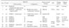
Among total 16 patients, 9 patients underwent corrective surgery and were followed-up for a median 72 months (range, 21 months to 11 years) (Tables 4, 5). Two patients who combined with cardiac anomaly and hydrocephalus, died without correction and 1 patient is currently awaiting corrective surgery. Remaining 4 patients were lost to follow-up.
Among 9 patients performed corrective surgery, 7 patients underwent Pena procedure as posterior sagittal anorectovaginourethroplasty (PSARVUR), and 3 patients of these required an additional abdominal approach for hysterectomy and colostomy dissection. Remaining 2 patients underwent PSARUR due to Mullerian agenesis.
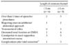
The median length of the common channels was 3.2 cm (range, 2 to 4.5 cm). The vaginal switch method was performed in 1 patient with a hemiuterus and hemivagina. The uterus and vagina of 1 patient had to be removed because of high located vagina and damage during dissection. Four patients underwent 3 procedures respectively, consisting of 1) a colostomy with a cystostomy or vaginostomy, 2) corrective surgery, and 3) colostomy restoration. Four patients required additional procedures after corrective surgery due to complications such as vesicovaginal fistula, urethral stone or urethral stricture. One patient combined with several associate abnormalities died of seizure in two years after corrective surgery.
Five patients with decreased renal function were assessed by vesicoureteral reflux and 99 mTc-dimercaptosuccinic acid tests. All live patients were able to do self-voiding. Two patients are trained by timed voiding, and since the rest two were below 3-year-old, it was unavailable to evaluate their voiding control. Only 1 patient was able to defecate without any supportive enema, and 8 patients needed enemas.
DISCUSSION
Persistent cloaca is a malformation in which the urinary, genital and digestive organs remain open toward the telomeric site of the perineum, mainly the rear of clitoris, through a common channel. This condition is due to abnormal development of the urogenital septum and is very rare, with a frequency of one per 50,000-125,000 newborns [1,2]. Since persistent cloaca has various clinical manifestations, it is not easy to diagnose by prenatal ultrasonography. Therefore, many of our patients were diagnosed as intraperitoneal cyst or hydronephrosis.
Since the patients with persistent cloaca can affect their mortality in neonatal period and the outcomes of corrective surgery and renal function, the appropriate diagnosis and treatment of these patients are very important [3-6]. A study of 361 patients with persistent cloaca identified 3 pitfalls in neonatal period management: 1) failure to recognize and manage hydrocolpos, 2) colostomy or vesicostomy problems and 3) misdiagnosis as imperforate anus with rectovaginal fistula and intersex [7]. The most important initial treatment is drainage of hydrocolpos, which can cause enlargement of the bladder. Subsequent cystostomy is recommended only when the bladder continues to be enlarged [7,8]. Other studies have found that 30% of children with persistent cloaca also have hydrocolpos [4,7], that suppression of trigone of bladder causes hydroureter and hydronephrosis and that infection of undrained hydrocolpos itself can result in perforation, sometimes accompanied by pyocolpos [9]. Since our study involved only a few patients, it is difficult to determine the efficacy of cystostomy and vaginostomy. Nevertheless, children who did not undergo drainage of hydrocolpos died of sepsis and complications, suggest that such drainage is essential. If self-voiding is possible, the need for cystostomy should be determined after assessing the degree of bladder enlargement at follow-up. Since we have not conduct vaginostomy only if both the vagina and bladder were enlarged, it is necessary to determine whether vaginostomy alone can cause bladder enlargement to disappear.
It is also most important to accurately determine the anatomical structure of each patient and to establish a treatment plan before cystostomy or vaginostomy. Urological defect is present in 68% children with persistent cloaca [6] with 33 to 50% having hemiuterus and hemivagina [6,10,11]. Various malformations were present in our patients, such as hemivagina, vaginal septum, blind pouch vagina, and Mullerian agenesis. Since hydrocolpos might not be drained appropriately, radiographic examination may be required before colostomy. A cystoscope can be used to measure the structure of the genital system and the length of the common channel, and a laparoscope can be used to diagnose hemiuterus and degree of enlargement.
Since 1982, corrective surgery has been performed using PSARVUR [6,12]. In addition, total urogenital mobilization can be performed by mobilizing the urogenital sinus from the pelvic cavity [13,14]. Using this method, it is easy to relocate the vagina down to the perineum when the length of the common channel is less than 3 cm, but, in patients with a common channel longer than 3 cm, there may be technical difficulties because the length may not be sufficient even after avulsion of both the vagina and urethra [15]. Other methods may be better in these patients, including vaginal switch and bowel replacement [9,11,16,17]. Although all patients underwent revision surgery using PSARVUR, several also required a laparotomy, increasing the number of patients with common channel ≥3 cm who are eligible for corrective surgery. Several patients also required additional operations due to complications after surgery. These children should be repeatedly examined for complications of the urinary system and renal function and require continuous bowel training. Since all patients had not reached puberty, we could not fully analyze the functions and results of the genital systems, suggesting the need for longer term follow-up.
In conclusion, three of twelve patients who had enlarged bladder or vagina at birth did not undergo cystostomy or vaginostomy at first operation, and they had earlier complications after surgery or required drainage tube insertion. Patients with anatomical malformations before colostomy, as well as hydrocolpos and bladder enlargement, require a vaginostomy with or without cystostomy to reduce complications. Follow-up is required in patients with hydrocolpos and bladder enlargement to determine whether vaginal drainage improves dilated bladder. Continuous long-term follow-up survey is required to determine the long-term results of corrective surgery.
 XML Download