

 ePub
ePub Citation
 Print
Print


INTRODUCTION
Inguinal hernia is among the most common problems that require surgical intervention for its treatment, and it is generally accepted that it should be electively repaired [1]. However, incarceration hernia sometimes happens and this has been reported to occur in 0.29 to 2.9% of all inguinal hernias [2]. Laparoscopic hernia repair, the extraperitnoeal approach and the transperitoneal approach have recently become popular procedures for not only bilateral inguinal hernia and recurrent hernia but also for unilateral inguinal hernia [3,4].
Several recent studies [5-8] have reported successful reduction and repair of chronic and/or even acute incarcerated inguinal hernias by the laparoscopic approach, yet this procedure is still controversial and strangulated inguinal hernias has traditionally been considered a contraindication for the laparoscopic approach. We conducted a retrospective analysis of the outcomes of 66 patients who underwent laparoscopic total extraperitoreal (TEP) repair for incarcerated inguinal hernias.
METHODS
A retrospective review and analysis of the medical records was implemented for the patients who were operated on using laparoscopic TEP repair from May 2002 to May 2010. Nine hundred forty five patients underwent laparoscopic TEP repair for unilateral inguinal hernias by a single surgeon; there were 66 patients who had incarcerated hernias and 879 patients who had non-incarcerated hernias. The assessed data included the outcomes, the demographic characteristics, the types and locations of the hernias, the operative time, the hospital stay, postoperative swelling, pain and recurrence, and the results of each group were compared.
The operative time was recorded from the time of skin incision to the time of skin closure. Post-operatively, we recorded the number of patients who needed more than two doses of analgesics for operative site pain. Swelling was defined as the presence of a palpable fluid collection or swelling over the operative site or scrotum during follow-up. The length of the hospital stay was defined as the total number of nights spent in the hospital after surgery. Most patients were discharged on the day after surgery. The patients were followed in the outpatient hernia clinic on a regular basis.
Operative technique
Laparoscopic TEP repairs were performed using the three-port technique. To create the pre-peritoneal space, a 12 mm transverse skin incision was made at the inferior edge of the umbilicus. The incision was carried down to the anterior rectus sheath. A small incision was then made on the anterior rectus sheath to expose the rectus abdominis muscle. A channel between the rectus muscle and the posterior sheath was created with blunt endopeanuts towards the symphysis pubis. A small tunnel was made between the rectus muscle and the peritoneum. The pre-peritoneal space was entered using a 45 degree telescope and insufflating CO2 gas at a maximum of 12 mmHg pressure. Finally, two accessory 5 mm ports were created: one port was 2 cm above the symphysis pubis in the midline and the other port was in the middle between the existing two ports.
After medial and lateral dissection, the cord was identified and separated from the hernia sac. For an incarcerated inguinal hernia, the contents were initially reduced from the sac into the preperitoneal space after the sac was opened and put back in the peritoneal cavity. The opened sac was closed using 5 mm endoclips (Ethicon Endo-Surgery Inc., Cincinnati, OH, USA); if the pneumoperitoneum was disturbed by the opened sac and this obscured the operative field, then decompression was done using a Veress needle. In some cases, external manual compression of the scrotum was needed for reduction of the contents; sometimes an additional incision was performed at the scrotum because the hernia contents could not be reduced by manual compression. If there was a band between the incarcerated omentum and the peri-intrascrotal area, then it was ligated and resected.
After handling the sac and peritonealization, a 13 × 8 cm surgipro mesh (Covidien, Mansfield, MA, USA) was placed and anchored with 5 mm spiral tacks (Tyco healthcare, Norwalk, CT, USA) in most patients, or it was left without anchoring.
In cases of an acute incarcerated hernia, after laparoscopic TEP repair we confirmed the viability of the incarcerated bowel via a converted umbilical port with an intraperitoneal approach; no additional incision was needed.
Statistical analysis
The operative time, the post-operative pain, the occurrence of swelling, the length of the hospital stay and the recurrence rates were compared by chi-square tests and independent t-tests. All the data collected from the database was analyzed using SPSS ver. 12.0 (SPSS Inc., Chicago, IL, USA). A P-value < 0.05 was considered to be significant.
RESULTS
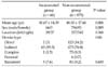
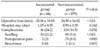
A total of 66 patients (all of the patients were men) with incarceration of inguinal hernias underwent laparoscopic TEP repair; an additional 879 patients underwent the same operation for unilateral non-incarcerated inguinal hernia during same period. The mean follow up was 45 months (range, 3 to 78 months). As Table 1 showed for the incarcerated group, the mean age was younger than that of the non-incarcerated group (41.67 ± 14.29 vs. 48.50 ± 17.66, P < 0.01), and most of the incarcerations occurred in indirect hernias (63 out of 66 cases, 95%). In Table 2, the mean operative time was longer for the incarcerated group (33.36 ± 18.83 minutes) than that for the non-incarcerated group (24.59 ± 16.92 minutes, P < 0.01). Postoperative swelling was more frequently observed in the incarcerated group (21.2%) than that in the non-incarcerated group (5.4%, P < 0.01). However, the hospital stay and postoperative pain showed no difference between both groups, and there was one recurrence in the non-incarcerated group, but this was not statistically significant.
There were 2 cases of acute incarcerated inguinal hernia in the incarcerated group. These 2 patients visited the hospital because of abdominal pain and inguinal area pain. Physical examination and imaging studies showed that there was no evidence of strangulation and manual reduction of the content failed, so an emergency operation was performed.
DISCUSSION
Laparoscopic TEP repair of inguinal hernia has become a more popular procedure because of the following reasons; less postoperative pain, faster recovery, a good cosmetic effect and a lower or similar recurrence rate as compared with that of conventional anterior repair [9]. After Watson et al. [10] initially described an emergency laparoscopic hernia repair associated with intestinal resection, several authors [5-8,11] have reported successful reduction and repair of incarcerated hernia with the laparoscopic approach, and even for acute incarcerated hernia. However, laparoscopic TEP repair for incarcerated inguinal hernia is still controversial, and traditionally the incarcerated inguinal hernias were repaired via open surgical procedure [12].
The laparoscopic approach for incarcerated inguinal hernia may have some problems; the peri-inguinal canal can be contaminated and this condition consequently can cause mesh infection, and the laparoscopic approach for reduction of the contents and repair of hernia requires a surgeon with great experience and the surgeon must be highly trained in laparoscopy [5,6]. However, the laparoscopic approach has an advantage over the open approach; there is no need for an additional incision for identifying whether or not the reduced contents are viable.
We applied laparoscopic TEP repair for incarcerated inguinal hernia after a learning curve (2 years of experience). In our series, only 2 cases of acute small bowel incarceration were included. Emergency operations were performed for these cases and fortunately the incarcerated contents were reduced spontaneously after anesthesia. In five cases, an additional incision was carried down on the scrotum because the contents could not be reduced with laparoscopic maneuvers. Further, there was no mesh infection, and this may be due to that most of the incarcerated hernias in our series were chronic and omental-incarcerated hernias.
In our results, postoperative swelling occurred more frequently in the incarcerated group than that in the non-incarcerated group. Chronic incarcerated hernia may have a large sac and more adhesion compared with non-incarcerated hernia, so sometimes extensive dissection was needed. Consequently, postoperative seroma, hematoma or cord swelling can frequently occur. Making the differentiation between cord swelling and real seroma after TEP is difficult if ultrasonography is not performed. However, most of the patients who underwent herniorraphy did not stay long in the hospital, and ultrasonography is not easily reachable study so only few reports were published previously about seroma or swelling after herniorrhaphy. The our previous studies have shown that most of the cases of swelling (83%) subsided spontaneously without any intervention, but other cases needed aspiration because of pain, discomfort or anxiety [13]. Even if none of our patients in the incarcerated group needed a drain, drain insertion in selected patient may be useful to decrease postoperative swelling if the hernia is the incarcerated type. Also, some authors [8,14] have suggested that selective drain insertion may be helpful to prevent mesh infection.
The operation time was longer because more time needed for reduction of the contents and repair of the sac in the incarcerated group, yet the operation time was acceptable (33 minutes). The length of the hospital stay and the postoperative pain were similar in both groups, and the recurrent rate was not different.
Laparoscopic TEP repair is not always possible for incarcerated inguinal hernia. It may be contraindicated when the incarcerated contents are suspected to be strangulated; the presence of skin redness or necrosis above the hernia site is a sign of this. In this situation, the surgeon has to be concerned about mesh infection, and delayed repair of a hernia during a second stage of treatment may be better [6].
This study has some limitations. Most of the incarceration contents were omentum, and only two small bowel incarceration cases were included. The subjects underwent laparoscopic TEP repair of non-incarcerated inguinal hernia, and they did not undergo the open conventional approach for incarcerated hernia. Although these are limitations, this study showed that laparoscopic TEP repair is safe and feasible for chronic incarcerated inguinal hernia. Also, in the case of acute incarceration, proper assessment of the viability of the hernia contents is crucial, and careful patient selection for applying TEP repair is very important.
In our opinion, laparoscopic TEP repair for a chronic or omental incarcerated inguinal hernia is safe, feasible and effective. However, for the cases of acute incarcerated hernia, more data is needed to confirm whether laparoscopic TEP repair is safe and effective, and especially for a suspected strangulation. Also, further investigation that will compare open hernia repair and laparoscopic hernia repair is needed to conclude which one is better for treating incarcerated inguinal hernia.
 XML Download