

 Citation
 Print
Print


INTRODUCTION
Acute gastrointestinal (GI) bleeding is a common emergency condition and an important cause of mortality. Furthermore, the GI bleeding source is not found in approximately 3-5% of patients by esophagogastroduodenoscopy (EGD) and colonoscopy. In these cases, most lesions responsible are found in the small bowel [1]. However, gastrointestinal bleeding originating in the small bowel is often difficult to diagnose and treat.
Hemangiomas originating from the small bowelare uncommon benign tumors, and may cause massive or occult GI bleeding. Although manytools can be used to diagnose tumors in the small intestine, hemangiomas with a small intestine origin are difficult to differentiate from other more common entities [2].
Recently, single incisional laparoscopic surgery has been utilized to treat various benign conditions in the abdomen [3]. Here, we report a case of jejunal polypoid hemangioma, causing recurrent GI bleeding and subsequent life-threatening anemia, which was treated using a single incisional laparoscopic approach.
CASE REPORT
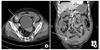
An 81-year-old female patient complaining of intermitt ent melena for 3 months, and of nausea and dizziness, which were aggravated just days before presentation, was found to have severe anemia. At presentation on emergency room, her initial hemoglobin and hematocrit levels were 4.7 g/dL and 15.8%, respectively. However, because her vital signs and performance status were stable, we went ahead the diagnostic evaluations with blood transfusions instead of the emergency operation. She had a medication history of hypertension and diabetes mellitus, but had not taken aspirin. EGD revealed only a 0.5 cm ulceration in stomach with no evidence of bleeding. Colonoscopic findings were unremarkable except for a 0.3 cm polyp in sigmoid colon, which also had no evidence of bleeding. An emergency abdominal CT scan was performed, and demonstrated a highly enhancing polypoid tumor in the distal ileum (Fig. 1). Diagnostic considerations included; adenocarcinoma or lymphoma, a polyp, a carcinoid tumor, or a vascular lesion. However, a GI contrast study failed to demonstrate any bleeding focus or mass lesion.
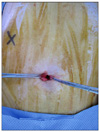
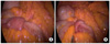
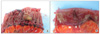
We decided to perform explorative laparotomy for diagnosis and treatment purposes. Surgery was performed using a single incisional laparoscopic approach with a surgical glove and a wound protector. The patient was placed in the supine position under general anesthesia with an endotracheal tube, and a 2-cm vertical transumbilical incision was made (Fig. 2) and the abdominal cavity accessed. A small Allexis Wound Retractor (Applied Medical, Rancho Santa Margarita, CA, USA) was placed, and a size 6 surgical glove was installed over the external ring. The thumb and middle finger of the glove were partially cut and 10-mm trocar for a videoscope and 5 mm trocar for a working device was placed and tied. When the intra-abdominal cavity was explored, an intussusceptum was found at the distal jejunal level (Fig. 3); no other abnormality was observed. The small bowel, including the intussusceptum, was taken out of the abdominal cavity, and segmental resection of the small bowel and end-to-end anastomosis by hand sewing were performed extracorporeally. The anastomosed small bowel was then placed in the abdominal cavity, and the wound was closed after saline irrigation. The resected mass was soft, pale brown, and 2 cm in size (Fig. 4), and was histologically confirmed to be a jejunal polypoid hemangioma (Fig. 5). The patient was discharged on postoperative day 7 without any complication.
DISCUSSION
Hemangiomas of the GI tract are uncommon, and account for only 0.05% of all intestinal neoplasms and 7 to 10% of all benign tumors of the small bowel. Ninety percent of hemangiomas are clinically evident, and present with symptoms, such as, acute or chronic GI hemorrhage, anemia, or obstruction, and rarely with platelet sequestration [4]. Other potentially serious complications of hemangiomas of the GI tract, such as, intussusception, small bowel obstruction, perforation, malabsorption, and bleeding from other sites of involvement, may also occur [5].
Bleeding is one of the symptoms associated with a small bowel neoplasm, and is usually occult and requires an extensive GI evaluation before a diagnosis is obtained. However, the diagnosis and localization of small bowel tumors remains a clinical challenge, because of the inaccessibility of this region to conventional diagnostic modalities. CT is frequently used as a front line tool for the evaluation of abdominal symptoms, especially in critically ill patients. CT scans show transluminal thickening of the wall of involved bowel loops with non-homogenous and persistent lesion contrast enhancement [5]. Double contrast studies demonstrate a nodular defect, which may change in configuration after compression or distension, which suggests a soft, possibly vascular tumor. The detection of this pathologic finding by double contrast study depends on the size of lesion and on the presence of active intestinal peristalsis [4,5].
Livengood and associates [6] described the feasibility of the angiographic localization of hemangioma of the small bowel. They performed angiography with methylene blue, which allowed lesions to be identified from an extraluminal vantage point. This method avoids the guesswork involved in transillumination and palpation for tumor localization during laparoscopy. In our case, we performed EGD, colonoscopy, an abdominal CT scan, and a double contrast study to indentify the bleeding focus, and with the exception of abdominal CT, these modalities did not indenty the problematic lesion.
We performed a single incisional laparoscopic exploration to localize and treat the jejunal tumor. Laparoscopic small bowel resection is an established technique and is performed by exteriorizing the diseased bowel segment and using traditional resection and anastomotic techniques [6]. Recently, multiple attempts have been made to reduce parietal trauma and visible scar formation even after laparoscopic surgery, and patient satisfaction has become a rapidly evolving issue, particularly in terms of single incisional laparoscopic surgery [3,7,8]. This issue reflects the importance of cosmesis and body image trauma associated with surgical procedures, and many surgeons have devised "scarless" surgical procedures using standard laparoscopic instruments. In the described case, standard laparoscopic instruments were used during the procedure.
Bleeding that originates from the small bowel presents challenges in terms of diagnosis, localization, and treatment. However, single incisional laparoscopic exploration may be helpful for localization and treatment purposes. In addition to its superior cosmetic results, a single incisional laparoscopic approach causes less morbidity by minimizing skin incisions. However, some bleeding lesions in the small bowel may be manifestations of a malignant process, and thus, it is essential that the surgeon has multiport laparoscopic skills, because these are vital for safe and effective single incisional laparoscopic surgery.

 XML Download