

 PDF
PDF ePub
ePub Citation
 Print
Print


Introduction
Although most mature cystic teratoma (MCT) occurs in the ovary, some cases of cystic teratoma of extra gonadal origin have been reported. It is usually found in neonates, and the ovary is most often implanted in the omentum.123 Ectopic ovary, used synonymously with accessory ovary, supernumerary ovary, or ovarian implant syndrome, is a rare gynecologic anomaly. Its etiology and true incidence is unknown. Ovarian auto amputation, especially occurring in ovaries with dermoid cysts, is a complication of ovarian torsion that may lead to formation of an ectopic ovary. Here, we present a rare case of an autoamputated ovary with MCT, and it is the first case of successful spontaneous pregnancy within seven months of resection.
Case Report
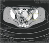
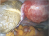
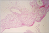
A 34-year-old woman, gravida 0, para 0 was referred to our clinic for presumed left ovarian tumor. Her past medical history was not significant; the patient had a history of chronic abdominal pain for two years. On pelvic examination, the uterus was normal in size. There was no tenderness in either adnexal region. Tumor markers were as follows, cancer antigen (CA) 125; 10.4 U/mL, CA 19-9; 2 U/mL. An ultrasonography examination was suggested the presence of 5.0 × 2.7 cm sized echogenic cyst in left ovary. A computed tomography (CT) scan was carried out and demonstrated a cystic mass measuring 5.7 × 3.1 cm at left ovary (Fig. 1). Laparoscopy was performed. Smooth, yellowish white mass was noted in the left adnexal region (Fig. 2). There was no ligamentous or direct connection with the pelvic organs, including the uterus, and there was no apparent blood supply to the tumor. The left ovary and tube were not found. The uterus and the right adnexa appeared normal. The mass was removed safely with blunt dissection. Histological examination revealed a typical MCT with adipose tissue and hair root sheaths. The cyst wall consisted of collagen fibers with marked infiltration of lymphocytes and histiocytes with calcification, which indicated ischemic and inflammatory changes (Fig. 3). These findings were consistent with a MCT within an autoamputated ovary.
Discussion
MCT is one of the most common types of ovarian tumors, with incidence ranging from 5% to 25% of all ovarian neoplasia.4 It is a germ cell neoplasm composed of various tissues, including tissues not normally found in the organ from which it arises. Embryologically, ovaries arise from the primordial germ cells that migrate from the wall of the yolk sac, along the dorsal mesentery, to the gonadal ridges.5 Theses totipotential cells may give rise to a variety of tissues originating from the three primitive germ cell layers. Dermoid cysts occur most commonly in the ovary, other favorite sites are the mediastinum, sacral region, and retroperitoneum. The incidence of parasitic dermoid cysts is 0.4% of all ovarian dermoid cysts. There are three proposed theories on the cause of these extragonadal sites: (1) primary dermoids originating from displace germ cells; (2) dermoids developing in a supernumerary ovary; and (3) autoamputation of an ovarian dermoid and implantation into an extra gonadal site.456 Autoamputation could result from the torsion of the pedicle, as torsion is reported to be the most frequent complication of ovarian teratomas, occurring in 16.1% of case.4 Torsion interferes with the blood supply, causing venous congestion and aseptic inflammation of the tumor wall. In acute torsion, the tumor undergoes necrosis and subsequent atrophy as a result of ischemia. In subacute or chronic torsion, the tumor may become adherent to adjacent structures, with a new collateral circulation formed. Infrequently, the tumor completely detaches from its pedicle, thus resulting in a parasitic dermoid cyst.7 This parasitic dermoid cyst may reimplant in adjacent structures and form a new blood supply. Thus torsion of ovary and its cystic contents may lead to development of a new ectopic ovary. The omentum is the main location for reimplantation of these parasitic dermoid cysts. The reason for the predilection for the omentum is because of its defensive role in intraabdominal inflammation, and adhesion formation, allowing the secondary implantation of the autoamputated ovary.8
Autoamputation is the most plausible mechanism for our patient whose history of chronic abdominal pain for two years. This pain most probably occurred after an ovarian torsion, thus resulting autoamputation. In contrast to other cases, it is suspected that the blood supply is cut off not long after autoamputation. In our case, the ultrasonography and CT suggested that the tumor might be MCT, but failed to demonstrate the exact localization of the tumor. However, it is suggested that the color flow Doppler may play an important role in the tumor localization.
In summary, our case report presents a patient with autoamputation of an ovary with MCT that was treated by laparoscopic surgery. One of the possible differential diagnosis is lipoleiomyoma of uterus, which contain lipid portion within mass that may lead to misdiagnosis of ovarian dermoid cyst.9 Some cases of gynecologic emergency may arise from postmenopausal women.10 Physician should keep in mind of this rare case when encountered postmenopausal women complaining of abrupt abdominal and pelvic pain. Furthermore, most of ovarian dermoid cyst can be treated by laparoscopic surgery with preservation of ovary, this kind of case only have consequence of oophorectomy which may lead to premature ovarian failure in patient with history of previous unilateral salpingo-oophorectomy.
 XML Download