

 PDF
PDF ePub
ePub Citation
 Print
Print


Introduction
Pyometra is collection of purulent material which occurs when there is interference with its normal drainage. It is an uncommon condition with incidence of 0.1 to 0.5% of all gynecological patients and 13.6% of elderly gynecological patients.1 Apart from genital tract malignancy and consequences of its treatment (radiotherapy), other benign conditions like endometrial polyp, fibroid, senile cervicitis puerperal infection and congenital cervical anomaly can lead to pyometra.2 Stenosis of cervical canal leading to accumulation of pus in the uterine cavity, degenerative or necrotic process in the uterine wall can lead to spontaneous perforation of pyometra. Spontaneous rupture of uterus is an extremely rare complication of pyometra.3 This case is being reported to present a spontaneous rupture of pyometra and generalized peritonitis managed by conservative surgery.
Case Report
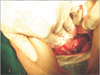
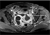
A 65-year postmenopausal lady, Para seven presented with foul smelling vaginal discharge associated with fever since fifteen days and pain in lower abdomen with mild distension of abdomen since five days. She was a known case of hypertension. On general examination her blood pressure was 180/120 mmHg. Her abdominal examination revealed distension of abdomen with tenderness in infraumblical region. Per speculum examination revealed normal cervix but frank pus was present which was sent for culture and sensitivity. On her pervaginal examination uterus was normal in size with mild tenderness. Ultrasonography and X ray abdomen in standing position were inconclusive. Magnetic resonance imaging (MRI) was done which revealed pyometra with uterine perforation in anterior wall with multiple loculated collection (Fig. 1), endometrium and cervix were normal. Her Pap smear was normal. Laparotomy was done. About 500 cc pus was drained and sent for culture and sensitivity. About 1 into 1 cm rent was present on anterior wall of uterus (Fig. 2). Both parametriums were thickened and inflammatory changes were present. Both fallopian tubes and ovaries were normal. Necrotic part of perforated area was excised and stitched. Peritoneal toileting was done. One intraabdominal drain was kept. Culture of the pus showed growth of gram positive cocci, sensitive to piperacillin. Patient was discharged on fifteenth post operative day.
Discussion
Pyometra usually present in elderly women. Nearly more than 50% of nonperforated pyometra patients are asymptomatic.4 Classical symptoms of these patients are purulent vaginal discharge, lower abdominal pain and postmenopausal bleeding. Non specific symptoms are common including vomiting, fever, nausea which leads to delayed diagnosis and eventually uterus gets perforated. Spontaneously perforated pyometra is difficult to diagnose preoperatively. The most frequent preoperative diagnosis are generalized peritonitis, pneumoperitoneum and perforated gastrointestinal (GI) tract.5 Correct diagnosis can only be made by laparotomy in most of the cases. Ultrasonography is the first investigation that has high sensitivity in assessing pyometra but has limited role in the diagnosis of perforated pyometra. Additional diagnostic radiographic evaluation use for acute abdomen is computed tomography (CT) scan and MRI.3 In our case-preoperative diagnosis of perforated pyometra was made by MRI. The treatment of ruptured pyometra is immediate laparotomy, peritoneal lavage, drainage, and/or simple hysterectomy.2 In most of the cases peritoneal cavity irrigation followed by total hysterectomy and bilateral oophorectomy is done. In present case patient was frail and uncontrolled hypertensive. On MRI endometrium and cervix were normal, Pap smear was also normal. We performed laparotomy followed by peritoneal toileting and repair of perforation. Patient had good recovery in postoperative period and discharged on fifteenth postoperative day. We want to highlight that preoperative diagnosis of perforated pyometra is absolutely essential. Patient care can be individualized and in selective patients of ruptured pyometra, conservative approach at surgery can be opted.
Conclusion
Although spontaneously perforated pyometra is rare, the condition must be borne in mind with regard to elderly women with acute abdominal pain. Preoperative diagnosis of perforated pyometra is absolutely essential because these patients are elderly, in poor general condition, and require prompt intervention. CT and MRI are diagnostic tools. Patient care can be individualized and in selective patients of ruptured pyometra, conservative approach at surgery can be opted.
 XML Download