

 PDF
PDF ePub
ePub Citation
 Print
Print


Introduction
Uterine myoma is one of the most common benign tumors in women and its prevalence rate has been known to be ranging 30% to 35%.1 In most cases of menopausal patients exhibiting uterine myoma, total hysterectomy is often recommended yet laparoscopic uterine myomectomy has been also widely adopted in cases where patients wish to conserve their uterus; in recent, a number of single port access (SPA) laparoscopic surgeries have been attempted in parallel with advances of minimal invasive laparoscopic operations.2,3,4,5 When compared to multiport access methods (using 3 or 4 ports access), it has been reported that there are not considerable differences between SPA against multiple ports access methods with regards to operation time, blood loss during operation, and degree of adhesion.6,7,8 In addition, recently concerns over diffuse intraperitoneal leiomyomatosis followed by morcellation (of uterine myoma) or uterine sarcoma are emerging hence it was proposed to perform a SPA method for morcellation thereby removing intraperitoneal residues more efficiently.9
A SPA laparoscopic myomectomy has its own benefits especially with regards to cosmetic aspect yet several issues exist such as hemostasis as well as delays in surgeries, compared to those of multi-port access methods. Thus, in the present study, we aimed to investigate and compare two suturing methods in a SPA laparoscopic myomectomy performed in our hospital.
Methods
1. Study subjects and methods
A total of 246 patients of laparoscopic multiple myomectomy were enrolled out of 869 patients underwent SPA laparoscopic operations in our hospital from August 2011 through December 2014. The present study was approved by the Institutional Review Board of the Pusan National University School of Medicine. All surgeries were done by seven surgeons and all patients records were retrospectively recorded/reviewed using their hospitalization records, outpatient records, and surgical records as follow: patients age, body mass index (BMI), pregnancy history, previous operation history, operative indication, uterine myoma sites, uterine myoma weight, numbers of uterine myoma, maximal sizes of dissected uterine myoma, intraperitoneal morcellation, suturing methods, sporadic uterine myomectomy, multiple uterine myomectomy, accompanied surgeries (e.g., ovarian surgery), operation time, degree of adhesion, amount of blood loss during operation, switching to multi ports access methods, exposure of intraperitoneal endometrium during operations, change in hemoglobin (before and after operation), change in hematocrit (before and after operation), transfusion, complications, and post-operative hospital stay. The operation time was defined as time took from navel incision to the point where skin incision was completed (and sutured). Changes in hemoglobin and hematocrit were estimated by measuring differences between the last measurement of hemoglobin and hematocrit (right before the operation) and the measurement done one day after the surgery. The amount of blood loss was calculated through the difference between the total quantities of infusedblood and the amount of blood used for washing. The complications indicated the status requires additional medical treatments potentially caused by the surgery; in this, post-operative fever lasted more than 5 days, urogenital system injuries, intestinal damages, intraperitoneal hematoma, transfusion, and reoperation were included. The post-operative hospital day was categorized into 7-days or shorter (which is considered to be general post-operative hospital days), 8-10 days, 11-14 days, and 15 days or longer due to complications.
2. Surgical methods
After providing general preparations, all patients were prescribed with prophylactic antibiotics 30 minutes in advance to their surgeries. General anesthesia was performed while maintaining the dorsal lithotomy position and a uterine manipulator was implemented after cervical expansion (KohColpotomizer, Colpo-PneumoOccluder, RUMI; Cooper Surgical, Trumbull, CT, USA). In order to perform the SPA, vertical navel incision, approximately 2 to 2.5 cm long, was made to reach the fascial plane which was removed after upper traction using the kelly. The intestinal adhesion was checked and then, the peritoneum was dissected followed by insertion of the Octo-PortTM to maintain the SPA. The rest of surgical procedures were identical with a general multi port access surgical method. Twelve mhg carbon dioxide was intraperitoneally infused and then a laparoscope was inserted (5 mm, 45 cm long; TelescopeTM, Karl Storz, Tuttlingen, Germany) to identify numbers and location of uterine myoma. All uterine myomas that were bigger than 2 cm were subjected to complete removal. In most surgeries, clamp instruments were 43 cm long and straight which is identical size of the laparoscope but some were flexible to bend if needed in their surgeries. To minimize blood loss in advance to performing myomectomy, a mixture of vasopressin (20 IU) and normal saline (1:80 dilution) was injected into subcutaneous uterine myoma and then uterine myomas were removed using a monopolar and bipolar electrocauteries. After removal, myometrium 8-shaped interrupt suture method was adopted in which uterine myomas were located adjacent to endometrium and then further proceeded with secondary suturing for myometrium and cervical layers; in contrast, if they were placed on outer myometrium, primary suturing was implemented for myometrium and cervical layers. In principle, secondary suturing was adopted for whom intended to get pregnant in future. In order to implement the secondary suturing technique, either 8-shaped interrupt suture method or continuous interlocking suture method was chosen and performed by a surgeon. Using a knot pusher, intracorporeal tie was done for the 8-shaped interrupt suture technique as shown in the Figure 1 and 2 utilizing polysorb 1-0 or 2-0. After myomectomy, uterine myomas, smaller than 3 cm, were retrieved via the navel hole without making another incision while morcellation of myomas was done through navel in a case where myomas were bigger than 3 cm. Although it was originally intended to prevent using an electronic morcellator (Gynecare, Somerville, NJ, USA) due to small fragments of uterine myoma, yet it was utilized in a limited way if unavoidable. A drainage tube was inserted via the navel incision after myomectomy and an uterotonics, either Carbetocin (Duratocin inj. 100 ug/1 mL) or Methylergometrine maleate (Eruvin inj. 0.2 mg/1 mL), was provided over 48 hours after the surgery.
Results
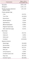
total of 246 patients were recruited and their age was ranging between 23 through 57 years old. The average age of patients was 39.94 years old (Table 1). The average BMI was 22.78 (± 3.35), while a total of 61 patients had their previous surgery history (22.3%). Sixteen and 26 patients had cesarean hysterectomy once (6.5%) or more than twice (10.6%), respectively whereas 2 patients had myomectomy previously. One and five patients previously underwent their uterine appendages and appendectomy, respectively while another patient had diagnostic laparoscopy. Lastly, other eight patients had a history of abdominal surgeries. When it comes to sites of uterine myomas, 14 cases, 2 cases, and 3 cases were placed on myometrium (5.7%), subserosal type (0.8%), submucous (1.2%), respectively yet most patients were having their myomas on multiple sites. Operative indications include excessive menstruation (59 patients, 24%), serious menstrual pain (30 patients, 12.2%), enlargement of uterine myoma size (14 patients, 5.7%), pelvic pain (3 patients, 1.2%), frequent urination (2 patients, 0.8%), accompanied ovarian tumors (24 patients, 9.8%), intraperitoneal large tumors (18 patients, 7.3%), and asymptomatic uterine myoma (86 patients, 35%). For patients with asymptomatic uterine myomas, surgery was determined in which 1) the size of tumor was bigger than 6 cm, 2) if a request was made by a patient with cancer phobia, and 3) a surgeon determined to do so.
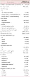
Surgical outcomes of the SPA laparoscopic myomectomy were summarized in the Table 2. The average numbers of removed uterine myomas were 2.31 (± 2.39; ranging from 1 to 15 myomas) excluding ones smaller than 1 cm. The size of uterine myoma was determined by the maximal axis length measured by tissue analyses and the average was 5.11 cm (± 2.11 cm). The electronic morcellator was used for one case yet morcellation was performed via single port using a surgical scalpel for most cases (i.e., 235 patients, 95.5%). Of these cases, small fragments of uterine myomas, possibly due to transformation thereof, were removed utilizing plastic bags (10 patients). One hundred seventy five patients underwent their myomectomy for multiple uterine myomas (71.1%) while 47 patients had myomectomy for single uterine myoma (47 patients, 19.1%). For 24 patients, uterine appendages were performed in parallel with myomectomy (24 patients, 9.8%). A total of 142 patients (57.7%) were representing mild adhesion with mesentery while 19 cases were either required to underwent cooperative treatment with department of surgery or suggested to have severe adhesion to intestine (7.7%).
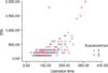
The average operation time was 107.68 minutes (± 50.61 minutes) and estimated blood loss was shown to be 263.67 mL (± 258.93 mL). The Pearson correlation coefficient and Spearman correlation coefficient between operation time and amount of blood loss were 0.706 and 0.674, respectively; both were statistically significant (Fig. 3). There were 2 cases that their surgical methods were switched to multiport access technique; of them, one patient was subjected to the abdominal section for the surgery. Three patients experienced the exposure of endometrium during operations (1.2%). The changes in hemoglobin and hematocrit were 1.34 (± 1.13) and 4.17 (± 3.24), respectively whereas blood transfusion was required for 29 patients (11.8%). Albeit three patients had intraperitoneal bleeding after their surgeries, re-operation was not needed for any case. General hospitalization days (i.e., less than 7 days) were required for most patients (i.e., 229 patients, 93.0%).
When it comes to suturing methods, primary suturing technique (i.e., 8-shaped interrupt suture) was performed for myometrium followed by secondary suturing technique, either 8-shaped interrupt suture method for myometrium and endometrium or continuous interlocking suture method; in the present study, we monitored differences in clinical outcomes between these secondary suturing techniques (Table 3).
Depending upon the secondary suturing techniques, a total of 160 patients had their interrupt suture while 86 patients underwent the continuous interlocking suture. In patients with the interrupt suture, the average uterine myomas removed was 2.31 (± 2.45) while it was 2.33 (± 2.28) in the patients with interlocking suture. The average sizes were 5.36 cm (± 1.94 cm) and 4.64 cm (± 2.33 cm) in the groups of interrupt suture and continuous interlocking suture, respectively. In the group of interrupt suture, the average size of myomas was bigger than those of continuous interlocking suture (5.36 ± 1.94 vs. 4.6 cm ± 2.33 cm, P < 0.05; Table 3), indicating that bigger size of uterine myomas were removed via the interrupt suture technique.
The averages of operation time were 100.50 minutes (± 42.09 minutes) and 121.04 minutes (± 61.56 minutes) in the groups of interrupt suture and continuous interlocking suture, respectively (P < 0.05; Table 3) while the amount of blood loss was 222.59 mL (± 144.94 mL) and 340.11 mL (± 380.62 mL), demonstrating less blood loss in the group with interrupt suture method. Changes in hemoglobin and hematocrit were 1.28 (± 1.23) and 1.44 (± 0.91), and 3.98 (± 3.47) and 4.52 (± 2.75) in the groups of interrupt suture and continuous interlocking suture, respectively yet no statistical significant was noted (Table 3).
The blood transfusion was made either before or after their surgery in 11 patients (6.9%) and 18 patients (20.9%) for the groups of interrupt suture and continuous interlocking suture, respectively. When it comes to postoperative complications six patients and three patients exhibited post-operative fever lasted more than 5 days (3.7%) and intraperitoneal bleeding (1.9%) in the interrupt suture group. In contrast, six patients complained post-operative fever lasted more than 5 days in the group of continuous interlocking suture yet no other complication was found. One hundred forty six patients were hospitalized less than 7 days in the group of interrupt suture (91.2%) while 83 patients did in the group of continuous interlocking suture (96.5%; Table 3).
Discussion
Since the first laparoscopic cholecystotomy was performed in 1987, significant advances in surgical instruments as well as clinical investigates have led to wide utilization of laparoscope in most surgeries. Compared to the abdominal section (also known as laparotomy), laparoscopic surgeries have been characterized by 1) considerable relief in postoperative pain, 2) less blood loss, 3) shorter hospitalization and recovery periods, 4) decrease in adhesion formation, and 5) preventing extensive scarring.6,7,8 The single-incision laparoscopic surgery (SILS) is one of the minimal invasive surgical methods which has been maximized with regards to their cosmetic benefits and is also known as laparoscopic single site surgery (LESS), SPA, and SILS. In 2001, the SPA laparoscopic appendectomy was firstly performed and then, laparoscopic cholecystectomy was implemented in 2007. Ever since then, many cases were reported to utilize the SPA technique in departments of urology, obstetrics and gynecology, and surgery. Currently extensive investigations are undergoing with regards to training hours for surgical techniques as well as developments of various surgical instruments (e.g., a single port, and instruments that are able to rotate and bend) in addition to conventional surgical techniques utilizing a straight instrument.10 Albeit these SPA laparoscopic surgeries are becoming more popular, the most serious concern over this method is a suturing technique and its usefulness because of difficulties to perform.
The SPA laparoscopic surgery may represent difficulties in hemostasis on myoma resection sites and suturing due to collisions between instruments compared to those of multi-port access methods. Widely utilized suturing methods include the 8-shaped interrupt suture method and continuous interlocking suture methods. The 8-shaped interrupt suture method uses a knot pusher for knot tying and repeats extracorporeal tying hence, only one site could be sutured at each time even though providing more sound tying; in addition, if a knot comes untied, the other sites may not be affected which is another benefit. Further, the interrupt suture method only utilizes one thread per each time thus more expensive and there is a more likelihood of adhesion due to extroversion of cervical layers. In contrast, the continuous interlocking suture method requires continuous suturing and intracorporeal tying; conventionally speaking, there are two specific techniques: 1) makes a loop on flat intraperitoneal site, let an instrument goes through the loop and then, and pulls the other side of thread through the loop and 2) wraps the thread using the other side of thread which is the typical method to do.10,11
In the present study, we demonstrated that the 8-shaped interrupt suture requires approximately 100.50 minutes (± 42.09 minutes) yet it was found to be significantly longer in the continuous interlocking suture (121.04 ± 61.56 minutes). In addition, when it comes to blood loss during surgeries, the interrupt suture had 222.59 mL of blood loss (± 144.94 mL) while the continuous interlocking suture represented 340.11 mL of blood loss (± 380.62 mL), demonstrating that the interrupt suture method results in smaller amount of blood loss which might be because it is relatively easier to perform and represents wider gaps compared to the continuous interlocking suture. In the interrupt suture method, fast suturing could be performed upon removal of uterine myomas thus blood loss might be reduced. Lastly, we also found positive correlations between operation time and blood loss (Pearson correlation coefficient: 0.706 and Spearman correlation coefficient: 0.674), supporting that ease of surgery (i.e., the 8-shaped interrupt suture) may significantly influence on the amount of blood loss. It is however, noted that changes in hemoglobin as well as hematocrit were not changed, which might be considered as statistical error biased by effects of blood transfusion made either before or after their surgeries.
On the other hand, the size of removed uterine myomas was 5.36 cm ± 1.94 cm in the patients underwent the interrupt suture method. In contrast it was found to be significantly smaller in the group of continuous interlocking suture method (4.6 cm ± 2.33 cm; P < 0.05). We demonstrated the positive associations between the size of uterine myomas and operation time; in results, the Pearson correlation coefficient and Spearman correlation coefficient were 0.245 and 0.245, respectively. Similarly, the associations between sizes of uterine myomas and blood loss were shown to be significant as demonstrated by the Pearson correlation coefficient (0.222) and Spearman correlation (0.259). These correlations indicate the statistical significance between the sizes of uterine myomas against operation time as well as blood loss. In the group of the interrupt suture method, the average size of uterine myomas, operation time, and the amount of blood loss were 5.36 cm, 100.50 minutes, and 222.59 mL, respectively. Meanwhile, these parameters were 4.64 cm, 121.04 minutes, and 340.11 mL, respectively in the group of continuous interlocking suture method; all were significantly different compared to the interrupt suture method. Such trends might be attributed to the faster and easier suturing technique (i.e., the interrupt suture method) chosen and implemented in which larger uterine myomas were observed thereby increasing operation time and blood loss. Albeit there were larger sizes of uterine myomas present, the average operation time and blood loss were significantly less in the group underwent the interrupt suture, clearly demonstrating it might be the better and faster suturing technique in regards to hemostasis.
Recently, the double tie method has been proposed, similar to conventional laparoscopic surgeries, via utilizing a surgical instrument that can be bending if needed. It is however, somewhat difficult and time-consuming. On the other hand, there is the barbed suture method which maintains tension through protuberances of threads although it accompanies risks of untangling as well as issues on their protuberances. Nonetheless, it was demonstrated that effective hemostasis as well as decrease in operation time could be achieved.12
Tam et al.13 compared a SPA laparoscopic method with a conventional laparoscopic surgery; the authors found no difference in clinical outcomes in which two crossing linear instruments were utilized. In other studies, it was further addressed that changes in position of z-axis direction, rotation of curbed instruments, and instruments with varying lengths may prevent crowding of instruments so that triangulation could be secured.13,14 In our study, we adopted curved instruments in early surgeries but straight instruments were utilized for approximately 20 patients after training. These straight instruments bear relatively stronger forces hence preventing frequent changes of instruments and shortening operation time.
Recently, concerns over diffuse intraperitoneal leiomyomatosis followed by morcellation (of uterine myoma) or uterine sarcoma are emerging thus it results increase in uterine sarcoma in menopausal women; these should not be overlooked in which laparoscopic myomectomy is performed for menopausal women.15 In the singly port access laparoscope, myomas could be removed without morcellation if they are smaller than the port size. Even if myomas are found to be larger than the single port, a surgical scalpel could be used to remove them without spreading of fragmentations through the port. In which a sarcoma is suspected, the Rab bag could be utilized and then proceed morcellation in order to prevent spreading myoma fragmentations. These may provide further usefulness and stability of laparoscopic myomectomy.
As opposed to the present study, a prospective investigation done by one single surgeon may provide more consistent and significant results in future; further, various investigations might be warranted with regards to effects of post-operative recovery, and degree of adhesion demonstrated after the surgery in order to shed further lights on advantages/disadvantages of the continuous interlocking suture method that could be overlooked in the study. As aforementioned, it is expected for the SPA uterine myomectomy to be utilized in various applications in parallel with advances in suturing instruments as well as development of various suturing methods.


 XML Download