

 PDF
PDF ePub
ePub Citation
 Print
Print


Introduction
Sclerosing stromal tumor (SST) is a rare benign ovarian neoplasm of the sex cord stromal category first delineated as a distinct ovarian sex cord stromal tumor entity in 1973 by Chalvardjian and Scully.1 Most patients afflicted with SST present with nonspecific symptoms related to an adnexal mass. The tumor, with rare exceptions, is hormonally inactive. Diagnosis of SST can be confirmed by postoperative pathologic examination. It is important to perform differential diagnoses of SST relative to other sex cord stromal tumors including fibroma, thecoma and lipoid cell tumors.2 It is distinguished from other ovarian stromal tumors by the production of collagen, a pseudolobular pattern, and it tends to occur in the second and third decades of patient life.3
Herein, we present two cases of SST occurred in postmenopausal women with brief review of literature.
Case Report
1. Case 1
A 63-year-old postmenopausal woman, gravida 5, para 2, experienced lower abdominal discomfort accompanied by a palpable abdominal mass for one month prior to her visit. Physical examination revealed a solid, non-tender tumor palpable up to the umbilicus level, and pelvic ultrasonography revealed a well-defined 14 cm sized heterogeneous, predominantly cystic pelvic mass with solid portions. A computed tomography (CT) scan of the abdomen and pelvis revealed a 15 cm sized tumor with an enhancing solid component, small amount of collected ascites and minimal peritoneal thickening. All clinical chemistry and tumor markers were below cut-off levels. Proceeding under the assumption that the finding was an ovarian torsion or cancer, the patient underwent a total abdominal hysterectomy including salpingo-oophorectomy, left external iliac lymph node sampling and washing cytology. We discovered an enlarged left ovary which had been torsioned clockwise twice and accompanied by necrotic changes. There was no evidence of metastases or peritoneal seeding.
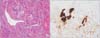
A gross examination revealed a 17.5 × 14.5 × 9.0 cm mass weighing 1248 g on the left ovary. Upon sectioning, the mass was found to be cystic with hemorrhagic fluid and a solid portion. Microscopically, the vessels contained a hemangiopericytoma-like staghorn pattern (Fig. 1A). The luteinized cells were strongly immunoreactive for inhibin alpha (Fig. 1B). The immunohistochemistry (IHC) results included inhibin α: positive, CD99: positive, desmin: positive, and actin: positive. Based on these histomorphologic findings, the diagnosis was SST.
2. Case 2
A 59-year-old postmenopausal woman, gravida 3, para 3, presented with complaints of abdominal pain with dysuria for 3 days prior to her visit. Upon clinical examination, a tender tumor was palpable up to the umbilicus level and was accompanied by left costovertebral angle (CVA) tenderness.
Ultrasonography revealed a 11.8 × 11.9 × 9.4 cm sized solid mass with a cystic component in the left adnexa. A CT scan of the abdomen and pelvis revealed a 14 cm sized solid-cystic mass and bowel wall thickening. The patient's serum cancer antigen 125 (CA-125) level was 37.9 U/mL (reference range < 35 U/mL). Proceeding with the assumption that this finding was a left adnexal mass, the patient underwent a total abdominal hysterectomy including both salpingo-oophorectomy and omental biopsy. A smooth, well-circumscribed, bosselated mass of approximately 15 cm in diameter was enucleated. The uterus, right ovary and bilateral fallopian tubes were normal in appearance.
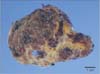
Gross examination revealed a 13.2 × 10.0 × 5.5 cm mass weighing 470.1 g on the left ovary. It was a cystic mass with a solid portion showing a diffuse hemorrhagic, variegated appearance. The cystic contents included dark brown serous fluid, blood clots and necrotic tissue. The solid components were white to yellow and fibrotic in appearance (Fig. 2).
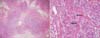
Microscopically evaluated, the solid area showed a pseudolobular pattern alternating between hypercellular and hypocellular areas (Fig. 3A). The cellular areas were composed of dual cell populations: collagen-producing spindle cells and rounded weak lutein cells (Fig. 3B). The results of the IHC assessment included Inhibin α: Focal positive, Calretinin: Positive, Smooth muscle actin: positive, desmin: focal weak positive and CD34: negative. The pathological diagnosis was SST of the left ovary.
Discussion
SST of the ovary is a distinct ovarian stromal tumor subtype.4 Ovarian SST most commonly occurs during the second to third decades of life at an average age of diagnosis of 27.5 years. More than 80% of SST cases occur in patients under 30 years of age.5 It is rare that SST is diagnosed in postmenopausal women, however, recently it has been reported that a 80-year-old woman had SST. It is far more difficult to diagnose SST in elderly women for many reasons; the incidence is much lower in elderly women, symptoms related to menstruation cycle can be hidden due to patients' menopause, and various common conditions can cause non-specific abdominal pain in senior populations.6
In children and adolescents, symptoms include premature menarche, menstrual irregularities, abdominal discomfort/pain, and rarely, ascites.6 In post-menarcheal females, SST presents with menstrual irregularities and/or an abdominal mass. In our cases, the patients presented with abdominal pain and a palpable mass. SST sizes vary from 1.5 to 20 cm in diameter.5
In patients with SST, serum CA-125 levels have been found to be either elevated or within reference ranges.7 The tumor in SST cases is usually hormonally inactive, although cases accompanied by irregular menses and genital bleeding have been reported. Peng et al.4, found 114 SST cases reported through 2003. In our two cases, we observed normal serum hormone levels with no clinical virilization. One patient's blood tumor marker was normal while the other was mildly elevated.
The sonographic findings associated with SST include a well-defined solid mass with hyperechoic honeycomb structures, which are also the characteristics of a mixed heterogenecity tumor without focal calcifications.8
It is difficult to diagnose before surgery by imaging studies. It used be diagnosed by pathological examination during or after surgery. A preoperative diagnosis of SST is possible based on magnetic resonance imaging (MRI) findings that demonstrate pseudolobulation, which consist of low-intensity nodules set against high-intensity stroma on T2-weighted images.9 The presence of tightly packed cellular areas associated with foci of sclerosis justifies the observed low density of these nodules on T2-weighted images. High-intensity areas found on the T2-weighted images correlated with poorly formed cellular tissue that was markedly edematous. However, the differentiation between SST and other stromal tumors and metastatic ovarian tumors based on MRI results needs further investigation. Upon the analysis of dynamic contrast enhanced images, the tumors revealed early peripheral enhancement with centripetal progression. Striking early enhancement reflects the cellular areas with their prominent vascular networks, and an area of prolonged enhancement in the inner portion of the mass represents the collagenous hypocellular area. These findings can be useful in differentiating SST from fibroma, as fibroma produces an absence of early enhancement and delayed accumulation of the contrast material.1011
Histologically, SST is characterized by cellular heterogeneity, prominent vasculature, and a pseudolobular structure composed of both cellular and hypocellular areas.12 The name "sclerosing stromal tumor" was proposed because the cellular portions of the tumor tend to undergo collagenous sclerosis. SST has occasionally been confused with massive ovarian edema and Krukenberg's tumor. The distinction between SST and Krukenberg's tumor can be made using IHC staining.4 SST is positive for desmin and smooth muscle action (SMA). Inhibin also has been shown to be positive and be a useful marker for ovarian sex cord stromal tumors.1314 In addition, both the cellular and edematous areas are positive for vascular endothelial growth factor. Other stromal tumors, i.e. thecoma and fibroma, tend to occur in the fifth and sixth decades of life of afflicted patients15 whereas almost 80 % of SSTs occur in women under 30 years of age.13
While most cases of SST have been reported to occur in the second and third decades of life, in this report, we present two cases of SST in postmenopausal women. We expect that this report will be helpful in the differential diagnosis of future SST cases that may occur in atypical patient populations.
 XML Download