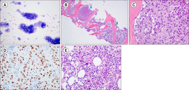

A 58-year-old man was diagnosed with essential thrombocythemia in 2010 and was managed with hydroxyurea and aspirin. In October 2014, he presented with right pleural effusion. Work-up revealed renal cell carcinoma metastasizing to the right pleura and lung parenchyma. Complete blood count (CBC) revealed white blood cells (WBC) 8.6×10⁹/L, hemoglobin 11.4 g/dL and platelets 294×103/µL. He underwent pleural decortication and right middle lobectomy, followed by left radical nephrectomy. In January 2015, sunitinib was started (after discontinuing hydroxyurea) resulting in initial tumor response. In May 2015, the cancer progressed and experimental trial was considered. At that point, CBC showed hemoglobin 9.6 g/dL, WBC 7.9×10⁹/L, platelets 446×103/µL. Bone marrow aspirate shows clusters of malignant cells (A, ×100). Bone marrow core biopsy revealed metastases (B, blue arrow, ×40) and residual myeloproliferative marrow (B, green arrow). Malignant cells with clear cytoplasm, large nuclei with prominent nucleoli and vacuoles are seen (C, ×400). PAX-8 staining was positive in carcinoma cells (D, ×400). Residual hypercellular marrow with dysplastic megakaryocytes with clustering in the residual marrow (E, ×400) is observed. This bone marrow biopsy thus reveals two simultaneously coexisting neoplastic conditions.


 PDF
PDF ePub
ePub Citation
 Print
Print


 XML Download