INTRODUCTION
Myeloid metaplasia (MM), also known as extramedullary hematopoiesis (EMH), refers to the physiopathological process in which hematopoietic progenitor cells differentiate into blood cells outside of the bone marrow [
1]. EMH, resulting from a compensatory mechanism for insufficient hematopoiesis, usually occurs in the spleen and the liver [
2]. Some authors have proposed that the main etiology of EMH are associated with hypoxia and acidic microenvironment [
345].
The splenic myeloid metaplasia (SMM) results from not only benign hematological diseases including autoimmune hemolytic anemias, immune thrombocytopenic purpura, and hemolytic uremic syndrome but also malignant disorder such as acute leukemia [
678]. SMM could occur secondary to filtration, insufficient production, or abnormal circulating cytokines or hematopoietic growth factors [
7]. In histological and immunohistochemical analysis, SMM usually show the lack of CD34, CD117, myeloperoxidase, and terminal deoxynucleotidyl transferase (TdT), with no evidence of clonal alteration in molecular and/or cytogenetic tests [
9].
The occurrence and its significance of SMM in warm autoimmune hemolytic anemia (wAIHA) are uncertain [
6]. The aim of this study is evaluating the frequency and clinical characteristics of SMM in patients with wAIHA, as well as comparing the differences in the response rates and disease-free survival (DFS) with those without SMM (categorized as individuals with splenic congestion). In addition to our previous report regarding the differences between primary and secondary AIHA [
10], we describe the clinical and pathological implications in our splenectomized patients in this paper.
Go to :

MATERIALS AND METHODS
Study design
We retrospectively analyzed 140 medical records of patients with wAIHA between January 1992 and December 2015 at the Hematology and Oncology Department of the Instituto Nacional de Ciencias Médicas y Nutrición Salvador Zubirán (INCMNSZ), a tertiary referral center in Mexico City.
Patients
Of these 140 patients, we included those fulfilled the following criteria: age >16 years, hemoglobin (Hb) ≤11 g/dL at diagnosis, demonstrable hemolysis (increased lactate dehydrogenase [LDH] and indirect bilirubin accompanied by haptoglobin), positive result in direct antiglobulin test (DAT) for IgG or IgG+C3d (mixed), and a history of therapeutic splenectomy as second-line treatment. The 36 patients fulfilled the inclusion criteria were evaluated for the presence of systemic lupus erythematosus (SLE) and antiphospholipid syndrome (APLS) with the following antibody panel: antinuclear antibodies (ANAs) >1:80 IU/mL, anti-double stranded DNA (dsDNA) antibodies >9.6 IU/mL, IgG anti-cardiolipin (ACL) antibodies or G phospholipids (GPL) >5.4 U, IgM ACL antibodies or M phospholipids (MPL) >4.4 U, IgG anti-apolipoprotein H antibodies (AAHAs) or anti-β2 glycoprotein I antibodies >2.5 U/mL, IgM AAHAs >3.4 U/mL, and positivity for the lupus anticoagulant (LAC) based on the parameters used by the Immunology and Rheumatology Department of our institution. We performed viral serology tests for the hepatitis B virus, hepatitis C virus, and human immunodeficiency virus in all the individuals included in this study. We excluded patients with incomplete hemolytic profiles at diagnosis, with incomplete clinical data, or with cold autoimmune hemolytic anemia (cAIHA). We considered the etiology as primary or idiopathic when we could not identify any autoimmune or malignant diseases provoking wAIHA. The patients were divided into two groups including patients with SMM or patients with splenic congestion (SC), depending on the histologic findings of spleen.
Histopathological analysis
The cases were reviewed by two experienced pathologists. Each spleen sample was stained with hematoxylin and eosin, and evaluated for the presence of extramedullary hematopoiesis. EMH was defined as when more than 10 groups of hematologic cells (erythroid, granulocytic series and megakaryocytes) were identified in the spleen samples.
Immunohistochemical reactions were performed using the automated Biocare system (Biocare Systems, Inc., CO, USA) and a standard streptavidin-biotin-peroxidase complex technique. The following markers were used: TdT (polyclonal DakoCytomation with 1:10 dilution) (Agilent Technologies, CA, USA), anti-myeloperoxidase (prediluted polyclonal DakoCytomation), glycophorin (polyclonal DakoCytomation with 1:200 dilution), CD34 (DakoCytomation, prediluted QBEND10 clone), and CD117 (Bio SB, C-kit with 1:50 dilution) (Bio SB, Inc., Ca, USA). The activity of the endogenous peroxidase was blocked with 3% hydrogen peroxide in methanol, and the activity of the endogenous biotin was blocked with avidin and biotin. The activity of peroxidase was developed with 3,3′-diamino-benzidine and it was counter-treated with hematoxylin.
Ethical approval
The study protocol was approved by the institutional review board of ethics and investigation of our institution with the reference number 1838.
Treatment
The patients received steroid regimen comprising three doses of IV methylprednisolone at 1 g/day, followed by 1 mg/kg/day of oral prednisone for 30 days, as first-line treatment. The second-line treatment was therapeutic splenectomy and the third-line treatment consisted of oral immunosuppressive agents like azathioprine (AZA), mycophenolate mofetil and cyclophosphamide (CYC). The response criteria were evaluated according to those of the
Gruppo Italiano Malattie Ematologiche dell'Adulto (GIMEMA) [
11]. A CR was considered with a Hb level of >12 g/dL and a normalization of all the hemolysis markers, while a partial response (PR) was considered with a Hb level of >10 g/dL or an increment of 2 g/dL over the basal Hb without the need for transfusions. Based on the above criteria, time to response was defined from the date of the beginning of treatment to the date of response (CR or PR). The absence of response or without response was defined as the lack of complete or partial response. Meanwhile, relapse was defined as a decrease in the Hb level or the appearance of hemolytic markers after the patient achieved a CR or PR.
Statistical analysis
The Kolmogorov-Smirnov test was used to determine the normality of the data. Descriptive data were presented as frequencies with percentages for categorical variables, and as medians with minimum and maximum values (range) for quantitative variables. Regarding the bivariate analysis, the Mann-Whitney U test was used for quantitative variables, and Fisher's exact test or Chi-square test were used for the categorical variables. Survival analysis was performed with the log-rank test and the Kaplan-Meier method. Patients with missing data were excluded when comparing the differences between two groups. All statistical analyses were considered as significant with a P<0.05 and performed with Stata 14 software (StataCorp LP, College Station, TX, USA).
Go to :

RESULTS
Patients
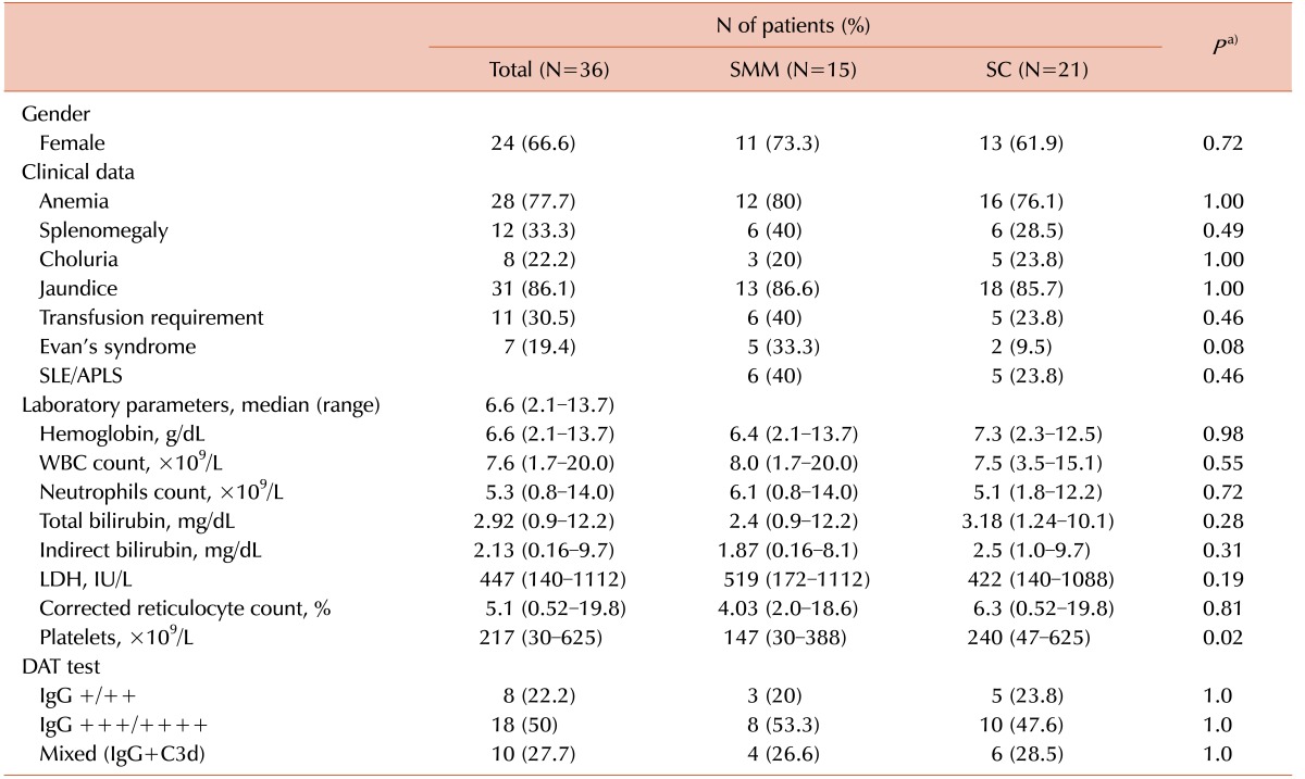
A total of 36 patients, including 25 (69.5%) patients with primary AIHA and 11 (30.5%) those with secondary AIHA, were included in the study. The demographic and clinical characteristics of patients are shown in
Table 1. Females were more common than males in both groups. In the histopathological analysis, 9 patients with primary AIHA and 6 patients with secondary AIHA showed the findings of SMM. The other 21 cases, including 15 patients with primary AIHA and 6 those with secondary AIHA, showed the findings of SC (
Fig. 1). The reasons for splenectomy were 25 cases (69.5%) of relapsed AIHA and 11 cases (30.5%) of steroid refractoriness. Among the 28 patients (77.7%) whose weight information of the resected spleens were available, the weight of spleen in SMM group was higher (median, 370 g, range, 98–1,105 g) than in SC group (median, 276 g, range, 145–830 g), albeit the difference was not statistically significant (
P=0.449).
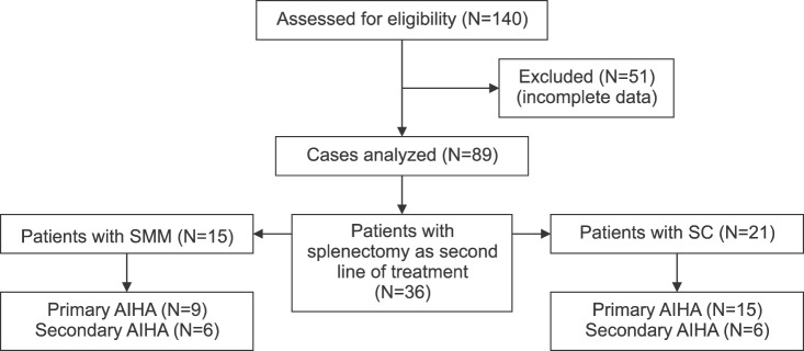 | Fig. 1Consolidated Standards of Reporting Trials (CONSORT) diagram of this study.
|
Table 1
Demographic and clinical characteristics of patients with wAIHA.

Clinical characteristics and laboratory parameters
When comparing clinical charateristics between two groups at clinical endpoint, no significant differences were shown in the transfusion requirements (40% in SMM vs. 23.8% in SC, P=0.46), the presence of splenomegaly (40% in SMM vs. 28.5% in SC, P=0.49), and the occurrence of Evans syndrome (33.3% in SMM vs. 9.5% in SC, P=0.08).
Among the laboratory parameters, the platelet counts were lower in SMM group (147×10
9/L) than in SC group (240×10
9/L) (
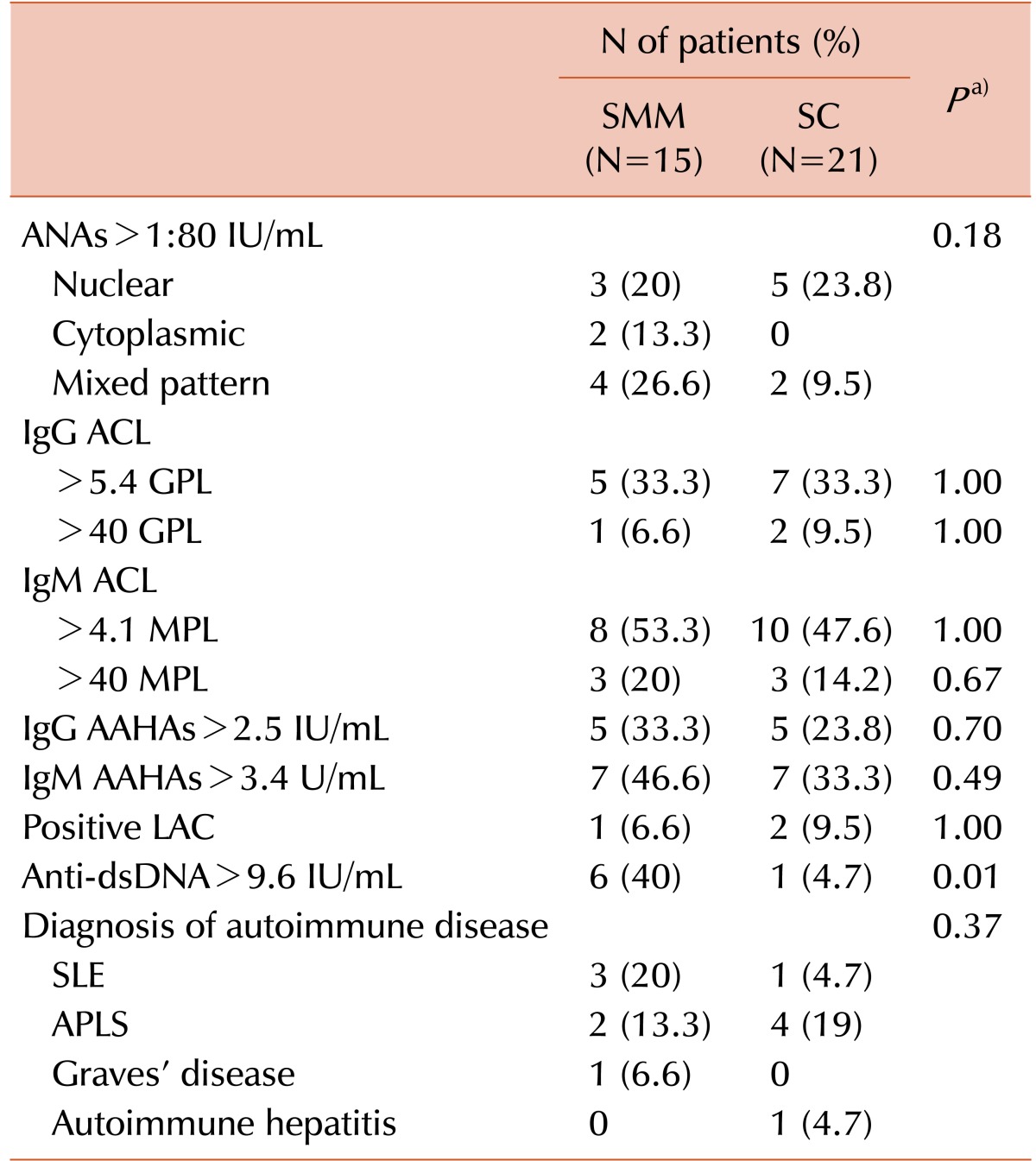
P=0.02). In addition, anti-dsDNA antibodies showed a statistically significant difference between the SMM group (40%) and the SC group (4.7%) (
P=0.01), whereas other rheumatologic profiles did not show any statistically significant differences as shown in
Table 2.
Table 2
Rheumatologic profile in patients with wAIHA.

Treatment response
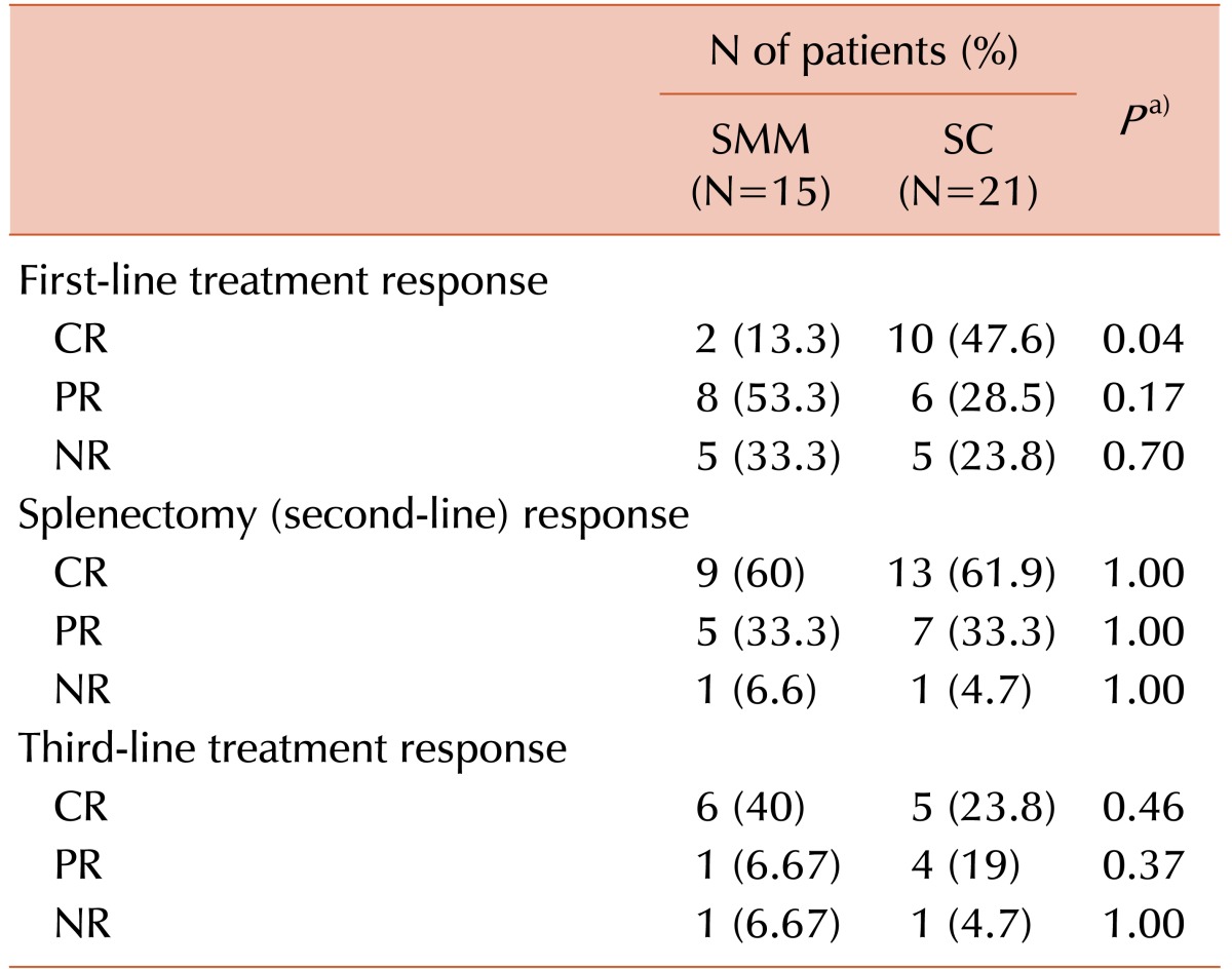
The treatment response to each phase of treatment is shown in
Table 3. The only CR of steroid therapy was statistically significant different (13.3% in SMM vs. 47.6% in SC) (
P=0.04). However, the therapeutic splenectomy and the immunosuppressive agents showed no statistically significant differences in treatment responses between two groups.
Table 3
Treatment responses between groups.

Although the median time from initial diagnosis to splenectomy was longer in the SMM group (3.9 mo) than in the SC group (4.6 mo), the difference did not reach statistical significance (
P=0.97). After splenectomy, the median DFS was 16.2 months in the SMM group, compared to 5.1 months in the SC group (
P=0.19) (
Fig. 2).
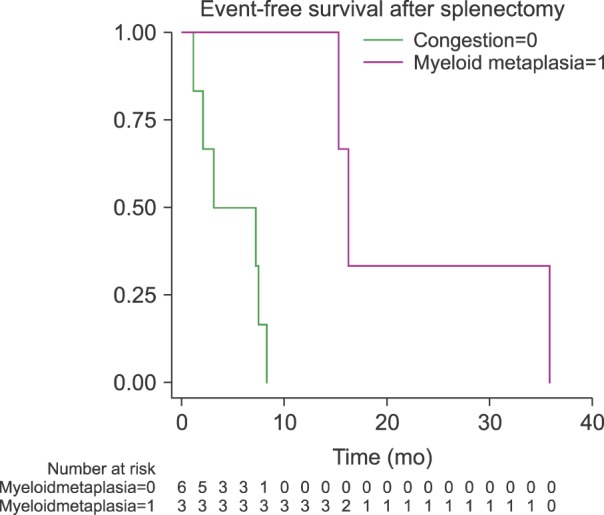 | Fig. 2Event-free survival after splenectomy. Event-free survival after splenectomy was 16.2 months in patients with MME versus 5.1 months in the splenic congestion group (P=0.19).
|
Univariate and multivariate analyses
In the univariate and multivariate analyses, achieving CR with the steroid regimen was a protective factor for the SMM group (odds ratio [OR] 0.3, 95% confidence interval [CI]: 0.03–0.94, P=0.03). The higher platlet count was also a protective factor in the SMM group (OR 0.99, 95% CI: 0.98–0.99, P=0.03); on the other hand, the presence of anti-dsDNA antibodies with titers above 9.6 IU/dL had a negative impact on this group (OR 2.76, 95% CI: 1.48–5.14, P<0.001).
Go to :

DISCUSSION
Overall, 15 patients (41.6%) showed the findings of SMM associated with wAIHA in our study. In few previous studies, the incidences of SMM in AIHA were highly variable (18–72%) [
121314]. In a retrospective study including 36 AIHA patients without lymphoproliferative disorder, 26 patients (72%) showed the finding of SMM [
14]. However, the authors of the study neither assess the clinical implications of this phenomenon nor further specify the AIHA cases as wAIHA or cAIHA. On the other hands, only 4 cases were associated AIHA in a multicenter study of United States including 80 SMM cases [
9]. Albeit not statistically significant in this study, females (73.3%) were predominant in the SMM group, consistent with the previous result (69.4%) [
14]. To the best of our knowledge, this is the first series that describes the presence of SMM associated to wAIHA secondary to SLE, APLS, and Evans syndrome.
In our study, we found no statistically significant differences in the hemolytic profile, the DAT results, and the requirement of transfusions between the SMM and SC groups. However, the platelet counts was a statistically significantly low in the SMM group compared with the SC group. Yam et al. [
6] found that patients with severe splenic hematoopoiesis had a platelet count below 50×10
9/L. To our own surprise, we did not find a significant difference regarding the presence of thrombocytopenia between the patients with and without Evans syndrome, and between those with and without SLE and/or APLS.
Regarding the treatment response, the SC group had a higher proportion of achieving CR to steroid than the SMM group (
P=0.04). This finding could be related to the chronic hemolysis in the SMM group, which could condition a poor response to the steroid regimen, as well as to a higher degree of disease, reflected by the larger hemosiderin deposits in the histological revision [
1214]. However, splenectomy and immunosuppressive agents did not show statistically significant difference in therapeutic response between both groups. Although splenectomy has been generally considered as a treatment modality for patients with steroid refractoriness or at relapse, the previous studies evaluating its use as a second-line treatment was not common [
111516]. To the best of our knowledge, this study is one of the largest scale series evaluating splenectomy as a standardized second-line treatment in patients with wAIHA. The overall responses higher than 90% of this study were superior to most of the previous results ranging from 38% to 75% of patients [
151718].
Although the difference was not statistically significant, patients belonging to the SMM group had a higher DFS median time after splenectomy when compared to the SC group. Since the phagocytic cells of the reticuloendothelial system, especially of the spleen, play a major role in hemolysis in patients with AIHA [
13], patients with SMM achieve longer DFS after splenectomy when compared to those without it, as reported in our results. This finding suggests that the main site of erythrocyte destruction in the SMM group is the spleen, whereas the erythrocytes in patients with SC are destroyed in other sites rather than spleen.
In our study, the presence of SMM in wAIHA had a correlation with elevated titers of anti-dsDNA antibodies. Among the overall 7 cases (19.4%) with increased anti-dsDNA antibody titer, 6 (85.7%) and 1 (14.3%) cases belong to the SMM group and the SC group, respctively (
P=0.01). Domiciano and Shinjo [
19] reported a higher prevalence of thrombocytopenia and positive anti-dsDNA antibodies in the SLE patients with AIHA than the those without AIHA. This result is consistent with the findings of the complete blood count and rheumatologic profile in our study. Similar observations have been described in a China study showing elevated anti-dsDNA titers in 37% SLE patients complicated with Evans syndrome (N=10/27) [
20]. Furthermore, no one has reported the negative impact of the anti-dsDNA antibodies for AIHA patients with SMM or EMH, except for the associations between IgG/IgM ACL antibodies and thrombocytopenia in patients with SLE/APLS [
212223],
The limitations of our study include the following aspects: its retrospective nature, the lack of long-term follow-up for both groups, and the use of conventional laboratory techniques. Despite this, our data suggest that patients with SMM and wAIHA have a different biological behavior from patients without SMM. To the best of our knowledge, this is the first study that describes the clinical impact of the histopathological spleen findings and their relation to the rheumatologic profile, specifically to the presence of anti-dsDNA antibodies. The role of the immune system in the development of SMM is yet to be defined, but we can suggest that the presence of anti-dsDNA antibodies play an important role in the development of SMM and in thrombocytopenia, as suggested by previous reports regarding patients with SLE/APLS.
Given its lower response rate to the steroid therapy and the association with anti-dsDNA antibodies, SMM should be of interest not only to the hematologist, but also to the rheumatologist. To further evaluate its frequency, clinical impact, hematologic manifestations, and influence in the development of rheumatologic disease, the long-term prospective studies with SMM patients are required to corroborate our data and understand of this phenomenon.
Go to :


 PDF
PDF ePub
ePub Citation
 Print
Print


 XML Download